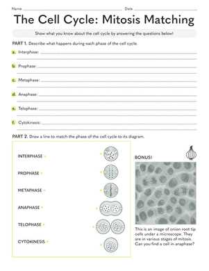
Cell division plays a crucial role in the growth and reproduction of living organisms. Understanding the process requires a clear grasp of how cells replicate and the stages involved in creating two identical daughter cells. It is essential to differentiate between each phase and the functions they serve in maintaining cellular integrity.
Educational exercises are commonly used to reinforce the knowledge of cell division. By identifying various components and their relationships, students can deepen their understanding and improve their ability to recall key concepts. These tasks challenge learners to match processes with their respective stages, enhancing critical thinking and retention.
To excel in these exercises, it’s important to focus on the specific actions occurring within each phase of cellular replication. Whether through diagrams, explanations, or practical examples, a comprehensive approach to the subject allows for better comprehension and long-term mastery.
Mitosis Matching Worksheet Answers
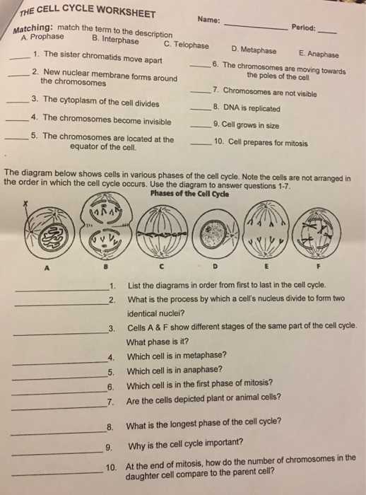
When studying the complex process of cellular replication, it is important to correctly identify and associate each step with its corresponding event. This involves understanding the distinct phases of cell division and how each phase contributes to the overall process. Proper recognition of each stage ensures that the key components and their roles are clearly understood, which is essential for grasping the fundamentals of cell biology.
Key Concepts for Correct Identification
During the division process, cells undergo a series of stages, each characterized by specific events that lead to the creation of two identical cells. Accurately identifying these stages is crucial for understanding the functionality of each phase. The challenge often lies in associating terms with their respective actions, such as the alignment of chromosomes or the separation of genetic material. Knowing these associations helps in mastering the topic and excelling in related exercises.
Common Mistakes and How to Avoid Them
One of the most frequent errors students encounter is confusing two stages with similar characteristics. For example, the final phase of division may be confused with the stage just prior to it, as both involve significant changes in the cell’s structure. To avoid such mistakes, it is important to focus on the specific details and changes that occur in each phase. Familiarizing oneself with diagrams and detailed descriptions can also aid in solidifying these concepts and preventing confusion during assessments.
Understanding the Stages of Mitosis
The process of cell division is critical for growth, repair, and reproduction in living organisms. It involves a series of well-defined steps, each with its specific function. Understanding these stages is essential for recognizing how cells replicate and ensure that the genetic material is evenly distributed to two daughter cells.
The Key Phases of Cell Division
Cell division consists of several stages, each marked by distinct events that prepare the cell for division and ensure that the process occurs efficiently. These phases can be divided into the following:
- Prophase: The initial phase where the genetic material condenses and becomes visible as chromosomes.
- Metaphase: Chromosomes align at the cell’s center, preparing for separation.
- Anaphase: Chromatids are pulled apart toward opposite poles of the cell.
- Telophase: The cell starts to divide, with new nuclear membranes forming around each set of chromosomes.
Why Understanding Each Stage is Important
Grasping the differences between each phase is vital for recognizing how cells accurately replicate their genetic material. A deep understanding of these stages provides clarity on the roles played by various cellular components, including the spindle fibers and centrosomes, which are crucial for chromosome movement.
By thoroughly understanding the sequence of events that occurs during this process, students and learners can more easily identify potential errors or issues that may arise during division, such as misalignment or improper chromosome separation.
Key Terms in Mitosis Process
Understanding the language of cell division requires familiarity with several key terms that describe the events and structures involved. Each term highlights a specific part of the process, from the initial preparation of the cell to the final separation of genetic material. Mastering these terms is essential for accurately describing the stages and understanding how each contributes to the overall process of cellular replication.
Chromosome: A thread-like structure composed of DNA and proteins that carry the genetic information. During division, chromosomes condense and become visible, ensuring that genetic material is evenly distributed.
Centrosome: A structure located near the cell’s nucleus that plays a crucial role in organizing the microtubules during division. It helps form the spindle apparatus that separates chromosomes.
Spindle fibers: Protein structures that extend from the centrosomes to the chromosomes. These fibers are responsible for guiding chromosomes during the separation process.
Chromatid: Each half of a duplicated chromosome, which is joined to its identical sister chromatid by a centromere. Chromatids are separated during the division process.
Centromere: The region of a chromosome where the two sister chromatids are joined. This structure plays a critical role in chromosome movement during cell division.
Cleavage furrow: A shallow groove that forms in the cell membrane during the final stage of division, marking the point where the cell will physically divide into two daughter cells.
Familiarity with these key terms provides a foundation for studying the intricate steps involved in the division process and supports deeper learning of cellular biology.
Common Mistakes in Mitosis Worksheets
When studying cell division, students often encounter challenges that can lead to common errors. These mistakes typically stem from confusion about the sequence of events, misinterpretation of diagrams, or misunderstanding key concepts. Identifying these common pitfalls can help learners avoid confusion and improve their understanding of the division process.
Frequent Errors Made by Students
There are several common mistakes that tend to occur when learners work on tasks related to cell division:
- Confusing the Phases: Many students mix up stages such as anaphase and telophase due to their similar characteristics. It’s important to focus on the distinct actions taking place in each phase.
- Misunderstanding Chromosome Behavior: A common mistake is incorrectly identifying the movement of chromosomes, especially during the separation of chromatids. Students often struggle with recognizing when the chromosomes are duplicated or when they’re splitting apart.
- Incorrect Labeling: Mislabeling structures such as the centromere, spindle fibers, or centrosome can lead to a poor understanding of their roles. Properly associating terms with the correct structures is essential.
- Overlooking Cell Plate Formation: In plant cells, students often forget to include the formation of the cell plate during the final stages of division. This key event is necessary for creating two daughter cells.
- Timing Issues: Some learners may have difficulty identifying when certain processes happen in the cycle, such as chromosome alignment during metaphase or nuclear membrane formation during telophase.
How to Avoid These Mistakes
To avoid these mistakes, it is important to study each phase carefully and to focus on the unique actions that distinguish them. Reviewing detailed diagrams, taking practice quizzes, and repeating the process of labeling and identifying structures can all help reinforce correct concepts and terminology.
How to Approach Mitosis Matching
When working on exercises related to cell division, it’s essential to break down the process and approach each part systematically. This not only helps in correctly identifying the stages but also improves your ability to recall specific events and structures. A strategic approach ensures a deeper understanding of the entire process and makes it easier to link the right terms with the corresponding stages of division.
Step-by-Step Approach for Success
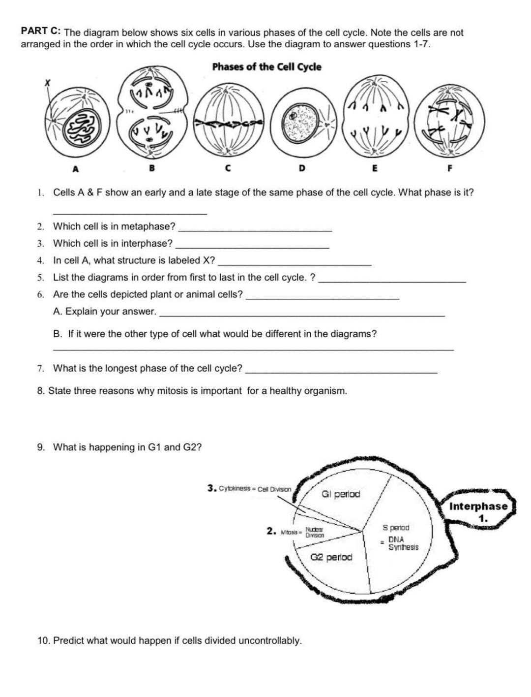
To effectively tackle these exercises, follow these key steps:
- Review the Stages: Familiarize yourself with the sequence of events. Knowing what happens in each stage of cell division will help you accurately connect the terms to their respective phases.
- Focus on Key Events: Pay attention to the critical actions that occur during each phase, such as chromosome alignment, separation, and the formation of new cell membranes. These events often serve as the distinguishing factors.
- Use Visual Aids: Diagrams and charts are incredibly helpful in visualizing the process. Try labeling these diagrams yourself to test your knowledge and make connections between the terms and structures.
- Take Notes: Keep a set of notes or a chart that lists the phases and their characteristics. This will serve as a quick reference during exercises and help reinforce the key points.
Staying Organized for Accuracy
To avoid confusion, it’s important to approach these tasks methodically. Begin by reviewing the options carefully, and eliminate terms that don’t match the phase in question. Then, double-check the characteristics of each phase before making your final choices. This process will reduce errors and improve your overall comprehension of the topic.
The Role of Chromosomes in Mitosis
Chromosomes play a central role in the process of cell division, ensuring that genetic material is accurately passed from one generation of cells to the next. These structures, which consist of tightly coiled DNA, undergo specific changes during the cell division process to allow for the proper distribution of genetic information. Understanding the role chromosomes play in this process is crucial for recognizing how cells maintain genetic stability.
During division, chromosomes undergo several important steps. They condense into visible structures, align at the cell’s center, and then separate to opposite ends of the cell. This careful movement ensures that each daughter cell receives an identical set of chromosomes, preserving the genetic information needed for normal cell function.
Chromosome Behavior During Cell Division
The behavior of chromosomes during cell division can be broken down into several key events:
| Event | Chromosome Action | Significance |
|---|---|---|
| Condensation | Chromosomes condense into visible structures | Ensures that genetic material is easier to manage during division |
| Alignment | Chromosomes line up at the cell’s equator | Prepares chromosomes for equal division between two daughter cells |
| Separation | Chromatids are pulled apart to opposite poles | Guarantees that each daughter cell receives a full set of genetic information |
| De-condensation | Chromosomes begin to unwind and return to a less condensed state | Prepares the genetic material for normal functioning in the new daughter cells |
By understanding the role and behavior of chromosomes, one can better appreciate how the cell ensures accurate replication and distribution of genetic material during division. This process is essential for the health and function of the organism as a whole.
Interpreting Mitosis Diagrams Effectively
Diagrams are a valuable tool for understanding the complex process of cell division. They provide a visual representation of the events that occur during this critical biological process, helping to clarify the sequence of actions that take place within the cell. However, interpreting these diagrams correctly requires careful attention to detail and a solid understanding of the underlying concepts.
Key Steps for Effective Interpretation
When analyzing diagrams related to cell division, it’s important to follow a systematic approach to ensure accurate interpretation:
- Identify Key Structures: Focus on the major components shown in the diagram, such as chromosomes, spindle fibers, centromeres, and the nuclear membrane. Understanding these structures is essential for recognizing the phase of division depicted.
- Recognize the Stages: Pay close attention to the specific features of each phase, such as chromosome condensation, alignment, and separation. Knowing these distinguishing factors will help you pinpoint the exact stage shown in the diagram.
- Understand the Sequence: Diagrams often show the stages in order. Be sure to recognize the progression from one phase to the next and understand how each stage leads to the next in the cycle.
- Look for Visual Cues: Key visual indicators, such as the position of chromosomes and the formation of the spindle apparatus, can help you identify what is happening at each step of the division process.
Common Mistakes to Avoid
While interpreting diagrams can be straightforward, there are some common mistakes that learners often make. Here are a few to watch out for:
- Confusing Stages: Many students may mistakenly identify a phase because they overlook key details like the arrangement of chromosomes or the presence of certain structures.
- Mislabeling Parts: Not recognizing the differences between similar structures (such as chromatids versus chromosomes) can lead to incorrect labeling and confusion.
- Overlooking Transitions: Some learners may fail to notice subtle transitions between stages, such as the gradual condensation of chromosomes or the formation of the nuclear envelope.
By focusing on these key factors, you can become more confident in interpreting diagrams and gain a clearer understanding of the complex events involved in cell division.
Visualizing the Mitosis Cycle
Understanding the series of events in cell division can be challenging, but visualizing the process makes it much easier to grasp. By breaking the division process into distinct phases, it becomes possible to see how each step contributes to the overall objective of producing two genetically identical cells. A visual representation of this cycle helps clarify the transitions and interactions that occur at each stage.
Breaking Down the Process into Phases
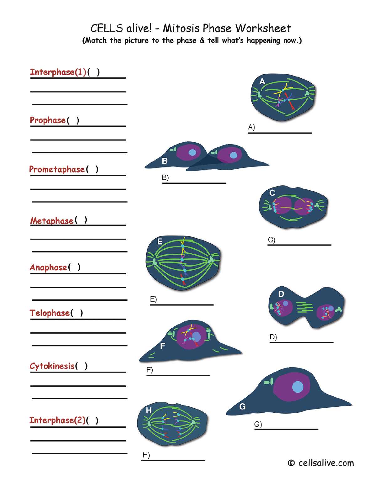
The cycle of cell division consists of several phases, each with unique characteristics. Visualizing these stages helps to connect the abstract concept of genetic material replication with tangible, observable actions. Here are the key phases that occur:
- Initial Preparation: The cell prepares for division by replicating its DNA and organizing the necessary machinery.
- Chromosome Alignment: Chromosomes align at the center of the cell, ensuring that each future daughter cell will receive a complete set of genetic material.
- Chromosome Separation: The chromosomes split into two chromatids, each moving toward opposite ends of the cell.
- Reformation of Structures: The cell’s membrane reforms, and the chromosomes unwind to restore the normal state of the cell.
Importance of Visualizing the Cycle
Seeing these stages in action allows students to grasp not only the sequence of events but also the precise actions involved. Diagrams that show these processes in order make it easier to understand how the cell ensures accurate genetic distribution. Visualizing the cycle also reinforces the idea of progression, from initial preparation to the final separation, solidifying the overall concept of cell division.
Why Mitosis is Essential for Growth
Cell division is fundamental to the growth and development of organisms. It allows living beings to increase in size, repair damaged tissues, and produce new cells to replace old or dead ones. Without this process, it would be impossible for organisms to grow, heal, or maintain proper function throughout their lifespan. Understanding how this division contributes to growth is key to appreciating its biological importance.
Facilitating Growth and Development
The process of cell replication enables an organism to expand by increasing the number of cells. As cells divide and multiply, tissues and organs grow, allowing for the proper functioning of the body. This is particularly crucial during the early stages of life, where rapid growth occurs, and also during the healing process after an injury.
- Development of New Tissue: As cells replicate, they form new tissue, supporting the formation of organs and systems necessary for life.
- Replacement of Old Cells: Cell division also replaces old or damaged cells, ensuring the integrity of tissues over time.
- Growth in Size: Increased cell numbers lead to an overall increase in the size of the organism, which is essential for survival and function.
Maintaining Health and Function
Cell division doesn’t just facilitate growth but is also essential for maintaining health. It allows for the constant renewal of skin cells, red blood cells, and other vital tissues. When this process functions properly, the body can maintain balance, repair injuries, and replace worn-out cells. Disruptions to this process can lead to serious health issues, including developmental disorders and tissue degeneration.
Challenges in Learning Mitosis Terminology
Understanding the vocabulary associated with cell division can be a significant hurdle for students. The terms used to describe the various stages and structures involved are often complex and can be easily confused, especially when there are similar words with slightly different meanings. Grasping the terminology is crucial for mastering the concepts, but it requires careful attention and practice to avoid misunderstandings.
Similar Terms with Different Meanings
One of the main challenges in learning the terminology related to cell division is distinguishing between similar terms. Many words describe processes or structures that sound alike but refer to different stages or components of the division cycle. For instance, terms like “chromatid” and “chromosome” can be confusing, as they both refer to genetic material, but in different forms during the division process.
- Chromosome vs. Chromatid: While both relate to the genetic material, a chromatid is one half of a duplicated chromosome, whereas a chromosome is the structure formed when the chromatids are joined together.
- Prophase vs. Metaphase: These terms describe two different stages of division, but they both involve significant events in chromosome movement, which can make them difficult to differentiate without a clear understanding of their specific roles.
- Centromere vs. Centrosome: These two terms sound similar but refer to different structures–centromeres are part of chromosomes, while centrosomes are involved in organizing the spindle fibers.
Overcoming the Complexity of Terms
To overcome these challenges, it’s essential to actively engage with the material, using visual aids, diagrams, and mnemonic devices to reinforce the definitions and relationships between terms. Repeated practice and active recall techniques can also help cement the terminology, making it easier to recall during exams or practical applications. In addition, contextualizing the terms in real-world examples or within the broader framework of biology can improve comprehension and retention.
How to Identify Each Mitosis Phase
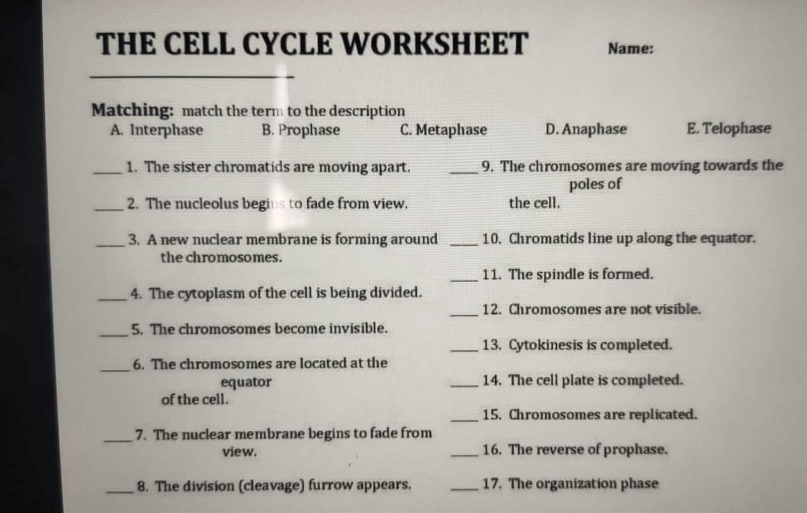
Recognizing the various stages of cell division can be challenging without a clear understanding of the key characteristics that define each phase. By focusing on specific features, such as the appearance and arrangement of chromosomes, spindle fibers, and other cellular structures, you can easily distinguish between each phase. These observations allow you to track the progress of cell division and understand how the process unfolds step by step.
To identify each stage, it’s important to focus on the following key visual cues:
- Initial Stage: Look for the cell with its full set of chromosomes, often as distinct, tightly packed structures. The nucleus may still be intact, and no division has yet occurred.
- Chromosome Alignment: In this phase, chromosomes align at the center of the cell. The spindle fibers should be visible, helping to guide the chromosomes into place.
- Separation of Chromosomes: During this phase, you will notice that the chromosomes split, with each half moving towards opposite ends of the cell, signaling the start of division.
- Final Division: In this phase, the cell’s membrane reforms around the separated chromosomes, and the cell splits into two, resulting in two distinct daughter cells.
By carefully examining these features and learning how they change from one phase to the next, you will be able to identify each stage with greater confidence and accuracy. Practice observing these transitions in diagrams or under a microscope to reinforce your understanding of the process.
Exploring Mitosis Worksheet Example Answers
Examining examples of cell division exercises can provide valuable insight into how the process of cell replication works. By reviewing these sample questions and solutions, students can gain a better understanding of the critical steps involved in cellular reproduction. These examples help reinforce theoretical knowledge while allowing learners to apply what they have studied to real-life scenarios. Each solution not only demonstrates the correct sequence of stages but also highlights common areas of confusion and how to overcome them.
When analyzing example problems, it’s helpful to break down each phase based on its unique characteristics. Here are a few aspects to focus on when reviewing these exercises:
- Identify Key Stages: Recognize the specific markers of each phase, such as chromosome condensation, alignment, and separation.
- Focus on Structures: Pay attention to the appearance of structures like spindle fibers, centromeres, and the nuclear envelope, as these are often the key indicators for each phase.
- Understand Timing: Some phases are very brief while others take longer. Understanding the relative duration of each step can provide additional context for the sequence of events.
By practicing with these examples, you will better understand how to interpret diagrams, identify the correct stage, and solve related problems with confidence. The more examples you work through, the more familiar you will become with the terminology and visual cues that define each stage of the process.
The Importance of Accurate Labeling
Proper identification and labeling of key elements in scientific diagrams are essential for understanding complex processes. Accurate labeling not only aids in reinforcing your knowledge but also ensures that the information you interpret or present is clear and correct. When it comes to cell division, being able to properly mark various stages, structures, and functions allows for a deeper comprehension of how each component contributes to the overall process.
Correctly labeling diagrams or exercises enhances your ability to:
- Visualize Complex Processes: Clear labeling allows you to connect theoretical knowledge with real-life examples, making abstract concepts more tangible.
- Ensure Clarity: Inaccurate or incomplete labels can lead to confusion, making it harder to understand the material or communicate your findings effectively.
- Improve Accuracy: When labels are correctly placed, it’s easier to follow the correct sequence of events and ensure a thorough understanding of the steps involved.
Labeling Key Structures in Diagrams
To effectively interpret cell division stages, it is essential to accurately label each part of the diagram, such as chromosomes, spindle fibers, and the cell membrane. Below is an example of a table showing the proper identification of these components during the division process:
| Structure | Function | Labeling Tips |
|---|---|---|
| Chromosomes | Contain genetic information and replicate during division | Label clearly, often depicted as X-shaped during certain phases |
| Spindle Fibers | Help move chromosomes during the division process | Indicate their position connecting the poles of the cell |
| Cell Membrane | Encloses the cell and eventually divides it into two daughter cells | Highlight the membrane especially during the final stages of separation |
Accurate labeling provides the foundation for analyzing and interpreting each phase, which ultimately enhances both understanding and retention of the subject matter. The clearer the labels, the better the comprehension of cell division and its stages.
Strategies for Solving Mitosis Matching Questions
When tasked with identifying and organizing key terms or phases in complex biological processes, effective strategies are essential for success. Understanding the sequence of events and relationships between components is critical to answering related questions accurately. By applying a methodical approach, you can ensure clarity and precision in your responses. Here are some strategies to help you navigate such tasks efficiently:
1. Break Down the Process Step by Step
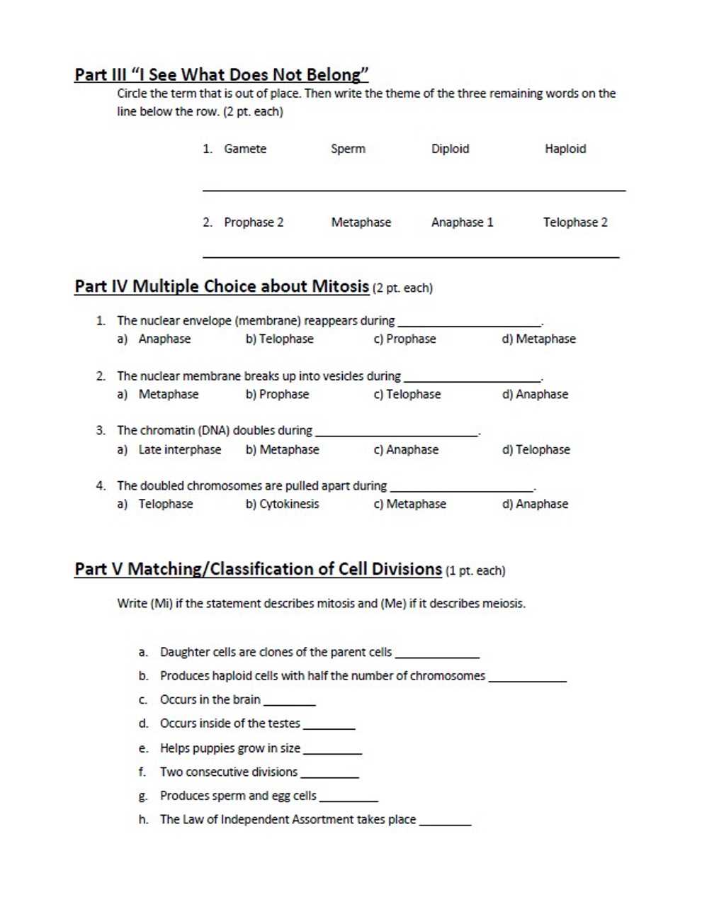
Understanding the flow of events can make it easier to match each concept to the correct stage or term. Here are some tips for breaking down the process:
- Understand the Phases: Familiarize yourself with the key stages involved and the transformations that occur at each step.
- Identify Key Terms: Recognize the terminology associated with each part of the process. Knowing the definitions will help in identifying what each term corresponds to in diagrams or descriptions.
- Use Visual Aids: Diagrams and flowcharts can help you visualize the process, making it easier to match terms with their correct positions.
2. Organize and Eliminate
Once you’ve broken down the terms and stages, the next step is to organize your approach and eliminate incorrect options. This strategy helps narrow down your choices:
- Group Similar Concepts: Group terms that relate to similar aspects of the process to avoid confusion later.
- Eliminate Incorrect Matches: If you can confidently identify the correct terms for some stages, eliminate these options from the list to focus on the remaining terms.
- Work in Sequence: Try to solve the questions in a logical order, from beginning to end, following the natural progression of events.
By adopting these strategies, you can effectively address challenges and ensure a more accurate and confident understanding of biological processes, leading to better results in matching tasks.
Common Errors in Cell Division Worksheets
When studying the intricate processes involved in cell division, students often encounter challenges that lead to common mistakes. These errors can arise from misunderstandings of key concepts, incorrect identification of stages, or confusion about the terminology used. Identifying these common pitfalls can help learners refine their knowledge and improve their accuracy in assessments. Below are some frequent errors that students make when working through exercises related to cell division:
1. Misunderstanding the Sequence of Events
One of the most common errors is a lack of clarity regarding the proper order of stages in the process. Students often mix up the phases, leading to incorrect answers. For example:
| Common Mistake | Explanation |
|---|---|
| Confusing Interphase with Prophase | Interphase is the longest phase, occurring before any visible changes in the cell, whereas prophase is the first actual stage of division. Mixing these stages can cause confusion. |
| Mislabeling Anaphase and Telophase | While anaphase involves the separation of chromosomes, telophase marks the final stages of division, where two distinct nuclei form. Confusing these two can lead to inaccuracies. |
2. Confusing Key Terms
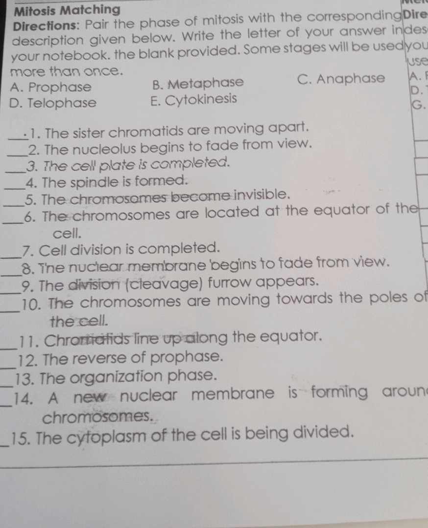
Another frequent error is using terms interchangeably, even when they describe distinct events or structures. For instance:
- Chromatid vs. Chromosome: Chromatids are individual strands of a chromosome that become visible during cell division, while a chromosome refers to the fully condensed form, which contains two chromatids.
- Cytokinesis vs. Telophase: Cytokinesis is the final process where the cytoplasm divides, whereas telophase focuses on the nuclear envelope reformation.
3. Incorrect Representation of Diagrams
Another common issue arises when students fail to accurately depict or interpret diagrams. These mistakes often stem from confusion about the visual markers for each phase or stage. Incorrectly labeling a diagram or misunderstanding the appearance of certain cell features can lead to errors. Key areas to watch for include:
- Correctly identifying spindle fibers
- Recognizing the formation of the cleavage furrow in cytokinesis
- Distinguishing between early and late stages of each phase
By being aware of these common mistakes, learners can focus their attention on areas where they might need further practice, leading to a more accurate understanding of cell division.
Reinforcing Mitosis Concepts for Students
For students to truly grasp the mechanisms of cellular division, it’s crucial to revisit and reinforce the foundational principles. This process is essential not only for academic success but also for fostering a deeper understanding of how life continues and evolves at the microscopic level. To enhance comprehension, students should be encouraged to actively engage with the material through various learning strategies, such as visual aids, hands-on activities, and collaborative discussions. Below are several effective methods for reinforcing key concepts related to cellular division:
Visual Aids and Diagrams
One of the most powerful tools in helping students solidify their understanding is the use of visual aids. Diagrams that accurately depict each phase of the division process can make abstract concepts more tangible. By working through these visuals, students can:
- Label key structures: Identifying and labeling chromosomes, spindle fibers, and cell membranes can help reinforce the structural changes that occur during division.
- Follow the sequence of events: Through the use of well-crafted illustrations, students can track the progression of each phase and understand the order in which events unfold.
- Compare different stages: Visual comparisons between stages help students recognize subtle changes in the cell’s appearance as it transitions through the various phases.
Interactive Activities and Practice
Engaging students in hands-on activities is another effective approach to reinforcing concepts. This can include:
- Model building: Using materials like clay or 3D printed models, students can physically construct and manipulate representations of cells, reinforcing their understanding of each stage’s structural changes.
- Quizzes and practice exercises: Regular, low-stakes quizzes allow students to check their comprehension and identify areas where they may need additional support.
- Group discussions: Encouraging collaborative learning allows students to articulate their understanding and hear different perspectives, which can deepen their grasp of the topic.
Real-World Applications
To further deepen understanding, it’s helpful to discuss the real-world significance of the process. Students can explore how cellular division impacts areas such as:
- Genetics and inheritance: Understanding how cells divide is key to grasping how traits are passed from one generation to the next.
- Medicine: Cellular division plays a crucial role in tissue repair, cancer growth, and the development of certain medical treatments.
By reinforcing these key concepts with a variety of interactive methods and real-world applications, students can develop a more thorough and lasting understanding of cellular division.