Understanding how medical professionals assess the interior of certain body parts is crucial for diagnosing various conditions. This examination is often carried out through advanced imaging techniques, allowing doctors to observe and analyze the condition of internal organs with precision.
Among these assessments, one of the most significant involves the inspection of a hollow organ responsible for storing waste. By using specialized instruments, experts can detect abnormalities and determine appropriate treatment strategies. These procedures are vital in identifying issues such as infections, blockages, or even more serious conditions like tumors.
In this section, we delve into the methods used for this type of inspection, including the tools, preparation steps, and potential risks involved. Additionally, we will explore how results from these examinations influence the overall care and management of patient health.
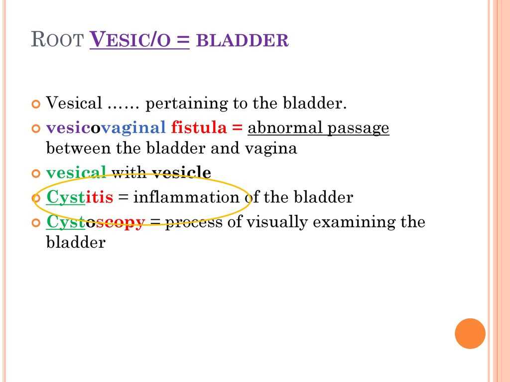
Process of Visually Examining the Urinary Bladder
When it comes to assessing the internal structure of hollow organs, precise methods are essential for diagnosing various health conditions. These procedures allow healthcare providers to inspect internal surfaces, detect potential issues, and determine appropriate treatment strategies.
In particular, one such technique involves using specialized tools to observe an organ involved in waste storage and elimination. Through this method, doctors can identify signs of inflammation, blockages, or other abnormalities that may not be detected through external examination alone.
Typically, the procedure is minimally invasive, using advanced imaging equipment that allows real-time visualization. This approach ensures that any irregularities are caught early, leading to better management and outcomes for patients. The examination is an important part of comprehensive care, especially when dealing with conditions affecting the flow or health of bodily fluids.
Overview of Bladder Visualization Techniques
In modern healthcare, various methods are employed to gain an in-depth look at internal organ structures. These techniques are crucial for diagnosing a range of conditions that might not be apparent through physical examination alone. Several approaches are used, each with its unique capabilities and benefits for detecting abnormalities and ensuring optimal care.
Among the most common techniques used to inspect hollow organs are the following:
- Cystoscopy: A minimally invasive method using a camera-equipped tube to directly observe the internal lining of the organ. This technique allows for high-resolution imaging and the ability to perform minor procedures simultaneously.
- Ultrasound Imaging: A non-invasive method that uses sound waves to produce images of internal organs. It is particularly useful for detecting changes in shape or size.
- CT Scan (Computed Tomography): A highly detailed imaging technique that combines multiple X-ray images to create cross-sectional views, providing clear images of internal structures.
- Magnetic Resonance Imaging (MRI): This imaging method uses magnetic fields and radio waves to produce detailed images of soft tissues, ideal for detecting abnormalities that might be missed by other methods.
Each of these techniques has specific advantages depending on the condition being assessed and the level of detail required. Some methods, like cystoscopy, allow for direct intervention, while others, such as ultrasound, offer a quick and non-invasive alternative for initial evaluations. Understanding the strengths and limitations of each approach is key to selecting the most effective method for diagnosing and monitoring health issues.
Why Visual Examination is Important
Evaluating internal organs plays a crucial role in diagnosing various medical conditions. By directly observing the interior structures, healthcare providers can detect abnormalities that are often hidden from routine physical assessments. Early detection is essential for preventing further complications and ensuring the right treatment is applied in a timely manner.
Key Reasons for Internal Assessment
There are several critical reasons why inspecting internal organs is important:
- Accurate Diagnosis: Direct visualization allows for a more precise identification of issues such as infections, growths, or blockages that might not be evident through other diagnostic methods.
- Minimally Invasive: Many modern techniques used for internal assessments are minimally invasive, reducing patient discomfort while providing essential information about health conditions.
- Real-Time Intervention: Some procedures allow healthcare professionals to intervene immediately if necessary, performing minor treatments or taking biopsies during the assessment.
- Prevention of Future Complications: Detecting abnormalities early enables effective treatment, reducing the risk of more severe issues developing over time.
Impact on Patient Care and Management
By utilizing visual assessment techniques, medical professionals can offer a more comprehensive care approach. These exams enhance the ability to monitor patients’ health, ensure accurate follow-up care, and tailor treatments based on detailed insights into their condition. This proactive approach helps in managing both acute and chronic conditions, ultimately improving patient outcomes and quality of life.
Different Methods for Bladder Inspection
There are several techniques used to inspect the interior of the organ responsible for waste storage, each offering unique advantages depending on the clinical situation. These methods range from non-invasive imaging tools to more direct approaches, enabling healthcare professionals to assess the health of the organ with great accuracy.
Each method provides different levels of detail and is chosen based on the symptoms, condition, and specific concerns of the patient. Some techniques allow for quick and broad evaluations, while others provide in-depth, real-time insight into the organ’s condition.
- Cystoscopy: This procedure involves inserting a thin tube with a camera into the organ, allowing doctors to directly view its lining. It is useful for detecting issues such as growths, inflammation, or blockages, and can also be used to take tissue samples or perform small interventions.
- Ultrasound: An external imaging technique that uses sound waves to create an image of the organ. It is non-invasive and particularly helpful in assessing the size, shape, and structure of the organ, though it may not provide as much detail as other methods.
- CT Scan: A highly detailed imaging technique that combines X-ray technology to create cross-sectional images of internal organs. It provides a comprehensive view of the organ’s structure, making it ideal for detecting larger abnormalities or growths.
- Magnetic Resonance Imaging (MRI): This non-invasive method uses magnetic fields and radio waves to produce high-resolution images, allowing doctors to evaluate soft tissue and identify subtle changes or abnormalities in the organ’s structure.
- Intravenous Pyelogram (IVP): This diagnostic test involves injecting a contrast dye into the bloodstream to highlight the organ and its surrounding structures on X-ray images. It is used primarily to detect issues like blockages or tumors.
Each of these methods plays a vital role in providing a clear picture of the organ’s health, helping to inform treatment decisions and ensure optimal patient care. The choice of technique is determined by factors such as the patient’s symptoms, medical history, and the suspected condition being investigated.
Role of Cystoscopy in Bladder Health

Cystoscopy plays a crucial role in assessing the health of the organ that stores waste, allowing doctors to gain direct insight into its condition. This technique provides detailed information that cannot always be obtained through external or non-invasive methods. By using a specialized instrument, healthcare providers can identify a variety of conditions that may affect the organ, ranging from infections to more serious issues such as tumors.
Benefits of Cystoscopy
This method offers several key advantages that make it a preferred choice for diagnosing conditions related to internal organ health:
| Benefit | Description |
|---|---|
| Direct Visualization | Provides real-time, high-resolution images of the internal lining, allowing doctors to spot abnormalities such as growths, lesions, or infections. |
| Minimally Invasive | The procedure is relatively simple and does not require major surgery, leading to shorter recovery times and less discomfort for patients. |
| Accurate Diagnosis | Enables precise diagnosis by directly observing the organ’s structure, which is essential for detecting hidden issues that may not show up on other tests. |
| Real-Time Treatment | Allows healthcare providers to perform minor procedures, such as removing small growths or taking tissue samples, during the same session. |
Common Conditions Diagnosed Using Cystoscopy
Cystoscopy is particularly effective in identifying various conditions that affect internal organs. Some of the most common include:
- Chronic infections
- Bladder stones or blockages
- Tumors or abnormal growths
- Inflammatory conditions such as interstitial cystitis
- Urethral strictures or narrowing
Thanks to its detailed imaging capabilities, cystoscopy allows for timely intervention and is an invaluable tool in maintaining overall health by detecting potential issues before they become more severe.
Preparing for a Bladder Examination
Preparation is essential for ensuring that internal assessments are as effective and accurate as possible. Proper preparation allows healthcare providers to obtain clear, detailed results, while also minimizing potential discomfort or complications for the patient. Understanding the steps involved in preparing for this type of examination can help reduce anxiety and contribute to a smoother experience.
Steps to Take Before the Procedure
Prior to undergoing the examination, there are several important steps to consider:
- Fasting or Dietary Restrictions: Depending on the method used, patients may be asked to avoid eating or drinking for several hours before the procedure. This ensures that the area of interest is clear and unobstructed.
- Hydration: In certain cases, staying hydrated is important to ensure proper imaging or to help with the flow of fluids during the examination.
- Medications: Informing the doctor about any medications being taken is crucial. Some drugs may need to be temporarily discontinued to avoid interference with the procedure or to ensure the safety of the patient.
- Clearance for Medical Conditions: Patients with specific health conditions may need special preparations, such as adjusting medications or receiving additional tests to ensure readiness for the procedure.
What to Expect During the Procedure
Understanding what will occur during the examination can help patients feel more at ease. The procedure is generally quick, and though it might involve some minor discomfort, it is typically well-tolerated. Medical professionals will guide patients through the steps, ensuring that everything is done with care and precision to achieve the best possible outcome.
Preparing adequately ensures that the assessment is effective, reduces the chances of complications, and contributes to a more positive experience overall.
Step-by-Step Guide to Bladder Inspection
When assessing internal organs related to waste storage, following a structured approach ensures that all necessary steps are followed for an accurate and effective evaluation. Each stage of the procedure is designed to provide clear, reliable results while minimizing discomfort for the patient. Understanding what to expect during each phase can help patients feel more prepared and confident.
Initial Preparations
Before beginning the examination, several preparatory steps must be taken to ensure everything is in place:
- Patient Consent: The patient must be fully informed about the procedure and provide consent before proceeding.
- Clearing the Area: The area of interest is cleansed to reduce the risk of infection and ensure optimal visibility during the examination.
- Positioning: The patient is positioned in a way that provides the healthcare provider with the best access to the target area. Comfort is important during this stage.
- Monitoring Vital Signs: Basic vital signs such as blood pressure and heart rate may be monitored to ensure the patient is stable before the procedure begins.
Performing the Examination
Once the preparatory steps are complete, the actual examination begins. This is typically done using specialized instruments or imaging techniques:
- Insertion of the Instrument: A thin, flexible tube with a camera is carefully inserted through the urethra to reach the target area. This allows the provider to view the interior directly.
- Inspection and Imaging: The healthcare professional inspects the lining and surrounding structures, often using additional tools such as light sources or imaging devices to capture detailed images or videos.
- Collection of Samples: If necessary, tissue samples or fluid may be taken for analysis, using tools that are passed through the same instrument.
- Completion of the Procedure: Once all required areas are assessed and samples are taken, the instrument is carefully removed. The patient is monitored as they recover from any minor discomfort.
Following these steps ensures a thorough and accurate evaluation, allowing medical professionals to diagnose conditions effectively and take appropriate action if needed. The method chosen will depend on the specific symptoms and health concerns being addressed.
Tools Used in Visual Bladder Exams
Various specialized instruments are essential for conducting internal assessments of the organ responsible for storing waste. These tools enable healthcare professionals to observe, diagnose, and even treat conditions within the internal area effectively. Different tools serve specific purposes, from providing clear images to facilitating sample collection.
Among the most commonly used tools are:
- Cystoscope: A flexible or rigid tube with a camera and light at the tip, used to view the interior of the organ. This tool allows doctors to directly observe the lining and surrounding structures.
- Ultrasound Probe: For non-invasive imaging, ultrasound technology can create detailed pictures of the area, helping professionals assess the shape and size of the organ.
- Biopsy Forceps: Used in conjunction with other tools, biopsy forceps allow for the collection of small tissue samples for further examination, helping to identify abnormalities such as tumors or infections.
- Light Source: A strong, focused light is often required during internal procedures to illuminate the target area, ensuring clear visibility and accurate assessments.
- Catheters: These are used to drain fluids or administer contrast material, which can enhance the clarity of the images captured during the procedure.
- Stents: In some cases, temporary stents may be placed to help maintain an open passage during or after the examination, especially if any obstructions are identified.
Each of these tools plays a crucial role in ensuring a thorough and accurate assessment. Depending on the specific requirements of the examination, healthcare providers may use one or a combination of these instruments to gather the necessary information and make informed diagnoses.
How Cystoscopic Imaging Works

Cystoscopic imaging is a technique used to capture detailed images of internal organs using specialized instruments. This method allows healthcare professionals to observe and assess the condition of tissues, identify potential issues, and guide further diagnostic or therapeutic actions. The process relies on advanced technology to provide a clear view of areas that are otherwise difficult to reach or see.
At the core of this method is a flexible tube, equipped with a camera and light, that is inserted into the body. The camera transmits real-time images to an external monitor, allowing the healthcare provider to examine the area in great detail. The light source illuminates the targeted region, ensuring that even the smallest abnormalities are visible during the procedure.
In some cases, additional imaging tools like ultrasound or contrast agents are used in conjunction with cystoscopy to enhance the clarity and precision of the images. These tools work together to provide a comprehensive view, helping professionals diagnose conditions with greater accuracy.
Ultimately, cystoscopic imaging plays a vital role in diagnosing and monitoring various conditions by offering an internal, high-resolution view, allowing for more informed medical decisions and improved patient care.
Common Conditions Detected by Visual Exams
Internal assessments play a crucial role in diagnosing a range of health conditions that affect the organ responsible for waste storage. These examinations can identify abnormalities that might not be apparent through external symptoms alone. By providing a direct view of the internal area, healthcare professionals can spot various conditions that require medical attention.
Some common issues detected during these procedures include:
- Infections: Inflammation or infection in the inner lining of the organ can be identified through changes in color, swelling, or tissue damage, indicating conditions like cystitis or other bacterial infections.
- Stones: Small, hard deposits can form within the organ, leading to blockages or discomfort. These stones are visible and can be evaluated for size and position to determine the best course of action.
- Growths or Tumors: Abnormal growths, such as polyps or cancerous tumors, may be detected as unusual masses within the organ. Early detection is critical for treatment and improving outcomes.
- Structural Abnormalities: Any congenital or acquired changes in the structure, such as scarring, diverticula, or deformities, can be observed and addressed promptly.
- Bleeding or Ulcers: Internal bleeding or open sores on the tissue can be identified, which might point to chronic conditions or acute injuries needing medical intervention.
These conditions, among others, can be effectively detected through these internal assessments, allowing for timely treatment and management of health issues. Early identification can significantly impact the outcome of medical treatments and recovery.
Risks and Benefits of Bladder Examination
Any medical procedure comes with its own set of advantages and potential drawbacks. Assessments of internal organs are no exception. While these procedures provide valuable insights into a patient’s health, it’s important to weigh the potential benefits against any associated risks to ensure informed decision-making. Understanding both sides helps individuals and healthcare providers make well-rounded choices when considering diagnostic or therapeutic options.
Benefits of Bladder Assessment
There are numerous benefits to performing internal assessments. These include:
- Early Detection: Identifying health issues in their early stages often leads to more effective treatment options and better outcomes. Conditions such as tumors, infections, or stones can be detected before they become more serious.
- Accurate Diagnosis: Direct visualization provides a clearer understanding of the condition, helping healthcare professionals make more precise diagnoses and reduce the need for invasive or costly tests.
- Targeted Treatment: By identifying the exact location and nature of a problem, the treatment can be more specific and less disruptive, leading to quicker recovery times.
Risks of Bladder Assessment
While the benefits are significant, there are some potential risks associated with these procedures. These include:
- Infection: Any procedure involving internal examination carries a risk of introducing bacteria, which can lead to infections, particularly if proper hygiene is not maintained.
- Discomfort: While most examinations are minimally invasive, some patients may experience discomfort or mild pain during or after the procedure, depending on the method used.
- Bleeding: In rare cases, minor bleeding can occur, especially if tissue is irritated or injured during the examination.
- False Positives: Although rare, there is a possibility of misinterpretation, where benign conditions might be mistaken for something more serious, leading to unnecessary procedures or treatments.
In conclusion, internal assessments are valuable tools in diagnosing health conditions, but like any medical procedure, they come with both benefits and risks. A thorough discussion with a healthcare provider can help to mitigate risks while maximizing the potential for early diagnosis and effective treatment.
Alternative Methods to Visual Bladder Inspection
While internal assessments offer direct observation, there are other diagnostic methods that can be used to evaluate the health of the organ responsible for waste storage. These alternatives can provide valuable insights, sometimes without the need for direct visualization. Depending on the patient’s condition and the specifics of the symptoms, these techniques may be preferable or used in conjunction with other assessments for a more comprehensive understanding.
Non-Invasive Imaging Techniques
Several non-invasive imaging technologies can be employed to observe the organ and detect potential issues:
- Ultrasound: This method uses sound waves to create real-time images of internal structures. It is commonly used to detect abnormalities such as stones, tumors, or fluid accumulation.
- CT Scan: A computed tomography scan provides detailed cross-sectional images of the internal organs, allowing for the detection of tumors, cysts, or other structural abnormalities.
- Magnetic Resonance Imaging (MRI): MRI uses powerful magnetic fields and radio waves to create high-resolution images. It is particularly useful for viewing soft tissues and detecting subtle changes or growths.
Functional Testing Methods
In some cases, functional tests may be employed to assess how the organ is working without visualizing its interior:
- Urinalysis: A simple test that can detect signs of infection, blood, or other markers indicating potential problems within the organ.
- Urodynamics: This method measures the pressure, flow, and volume of urine, helping to assess how well the organ stores and releases waste.
- Blood Tests: Blood tests can detect markers of infection, inflammation, or kidney function, indirectly indicating potential problems with the internal organ.
Each of these alternative methods has its own strengths and limitations, and often, a combination of techniques is used to form a clearer picture of the patient’s condition. These methods are valuable tools that can be employed when direct visualization is not possible, offering safer and more comfortable options for many patients.
Understanding the Cystoscope and Its Use
A cystoscope is a specialized medical instrument designed to provide a detailed view of the internal structures responsible for waste storage and excretion. It is an essential tool that allows healthcare professionals to directly inspect areas of concern, helping in the diagnosis of a variety of conditions affecting the lower abdomen and related organs. Through its use, doctors can detect abnormalities, infections, or signs of disease that might not be visible through standard imaging techniques.
The device consists of a long, flexible tube equipped with a light source and camera, enabling clear, real-time images of the inner structures. By inserting the cystoscope into the body through natural openings, the practitioner can gain access to the specific area of interest without the need for invasive surgery. This makes the procedure less traumatic for the patient while providing valuable diagnostic information.
Types of Cystoscopes:
- Flexible Cystoscope: This type is commonly used for routine assessments. It is lighter and more flexible, providing greater comfort during insertion and allowing easier movement to view different areas.
- Rigid Cystoscope: The rigid variety is typically used for more detailed examinations or procedures that require precision. Though less flexible, it is often used when larger instruments or surgical tools need to be passed through the scope.
Applications of Cystoscopy:
- Diagnosis: It helps identify conditions like infections, tumors, stones, or structural anomalies.
- Treatment: Besides diagnostics, cystoscopes are also used in minor surgeries, such as removing stones or taking biopsies.
- Monitoring: Patients with ongoing conditions may undergo repeated cystoscopies to monitor progress or changes in their condition over time.
The cystoscope plays a critical role in modern urological care, offering a minimally invasive way to assess and treat a range of conditions with high precision and minimal risk to the patient.
Interpreting Results from Bladder Visualizations
After capturing images or video footage of the internal structures, healthcare professionals must carefully analyze the findings to determine whether there are any abnormalities or issues present. These visual assessments offer crucial insights into the condition of the organs involved in waste storage and excretion. The images provide a direct view of potential issues, ranging from infections to structural changes, allowing for accurate diagnosis and effective treatment planning.
Understanding the results requires a combination of technical knowledge and clinical experience. Specialists will look for signs of inflammation, growths, blockages, or irregularities in shape or function. In many cases, further tests or procedures may be recommended based on initial findings. Below are some common factors considered during interpretation:
Key Aspects to Consider
- Color Changes: Variations in color may indicate inflammation, infection, or bleeding. Yellowish or reddish tones might suggest infection, while darker shades could signal other conditions.
- Shape and Size: Any irregularities in size or shape can point to conditions like tumors, cysts, or structural abnormalities. A healthy organ should have a smooth, consistent appearance.
- Presence of Foreign Objects: Small stones, clots, or other foreign materials can obstruct function and may require removal or additional treatment.
Common Findings and Their Implications
| Condition | Possible Implications |
|---|---|
| Infection | Redness, swelling, and discharge may indicate an infection that requires antibiotics or other treatments. |
| Growths or Tumors | Any abnormal masses or lesions could suggest the need for further testing, such as biopsies, to rule out cancer. |
| Blockages | Obstructions in the passageways could cause pain, discomfort, or difficulty with urination. Removal or surgical intervention might be necessary. |
| Congenital Abnormalities | Structural differences from birth may need to be monitored for potential complications, though they might not always require treatment. |
Accurate interpretation of these findings is essential for determining the right course of action. Depending on the diagnosis, treatments can vary from medications to surgical procedures, and a thorough understanding of the visualization results guides healthcare providers in making the best decisions for the patient’s care.
How to Maintain Bladder Health Post-Examination
After undergoing an internal inspection, it is important to adopt lifestyle habits that promote ongoing health and well-being of the organs involved in waste storage and elimination. Taking proactive steps can help prevent complications, support recovery, and ensure long-term optimal function. Maintaining a healthy routine will also help reduce the risk of recurring issues or further investigations.
There are several key strategies that can contribute to sustaining health after such a procedure. These practices include staying hydrated, managing nutrition, and monitoring any potential symptoms. Below are some important guidelines to follow:
Hydration and Diet
- Stay Hydrated: Drinking plenty of water helps flush out toxins and maintain the function of the waste-eliminating organs. Aim for 6-8 cups of water daily, unless otherwise advised by your healthcare provider.
- Avoid Irritants: Some foods and drinks, such as caffeine, alcohol, and acidic beverages, can irritate the internal organs. Reducing consumption of these items can reduce discomfort and prevent inflammation.
- Eat Fiber-Rich Foods: A diet high in fiber can promote better digestion and prevent constipation, which in turn supports better organ health.
Managing Symptoms and Monitoring Changes
- Monitor for Symptoms: If you notice any changes, such as discomfort during urination, unusual frequency, or blood in urine, consult with your doctor immediately.
- Avoid Overexertion: Engage in regular, low-impact exercises rather than straining physical activity that could cause stress or irritation to the region.
- Regular Follow-Ups: Attend scheduled check-ups to monitor your recovery progress and address any emerging concerns as early as possible.
Prevention Tips for Long-Term Health
- Maintain a Healthy Weight: Excess weight can put pressure on the organs and increase the risk of complications. Regular physical activity and a balanced diet can help maintain a healthy weight.
- Practice Good Hygiene: Keeping the area clean and free of infections is essential. Ensure proper hygiene practices to avoid irritation or bacterial growth.
- Don’t Hold Urine: Regularly emptying the bladder is important. Holding urine for extended periods can lead to overextension and potential infections.
By following these preventive measures and staying vigilant, you can help maintain the health and function of the organs responsible for waste elimination. Continued attention to your overall well-being and adhering to professional advice will support a healthier and more comfortable lifestyle.
Recent Advances in Bladder Visualization Technology
Recent innovations in technology have significantly improved how internal organ assessments are conducted, especially those related to waste storage and elimination. The development of advanced imaging techniques and tools has made procedures less invasive, more accurate, and faster. These innovations provide healthcare providers with clearer views and more precise information, which plays a crucial role in diagnosing and treating various conditions affecting the internal organs.
With the continuous progress in medical imaging, several cutting-edge technologies have emerged. These methods offer greater clarity and efficiency, providing both patients and doctors with enhanced confidence in diagnostic results. Below are some of the most notable advancements:
High-Definition Cystoscopes
High-definition cystoscopes have dramatically improved the clarity of internal views, allowing for more detailed and accurate visualization. These advanced devices are equipped with enhanced resolution, which makes it easier for medical professionals to identify abnormalities, lesions, and other irregularities. They provide a clearer, real-time picture, reducing the need for repeat procedures and minimizing risks.
Fluorescence Imaging
Fluorescence imaging is a promising technique that helps highlight specific areas of concern within the internal structures. By using special dyes, this technology can target abnormal tissues, making it easier to identify conditions like cancer or inflammation. It allows for more precise detection and improved outcomes, particularly in cases where early intervention is key to effective treatment.
3D Imaging and Virtual Reality
Another significant advancement involves the use of 3D imaging and virtual reality (VR) technologies. These methods allow for a comprehensive, multidimensional view of internal organs. With 3D visualization, healthcare providers can rotate and zoom in on images, offering a more detailed and interactive approach to diagnosis. VR integration is particularly useful for surgical planning, enabling doctors to better understand the patient’s anatomy before performing any interventions.
As technology continues to evolve, the future of internal organ assessments looks promising. These new tools and methods are enhancing diagnostic accuracy, reducing patient discomfort, and opening up new possibilities for non-invasive treatments. By embracing these advances, medical professionals can improve patient outcomes while providing a more comfortable experience throughout diagnostic procedures.
Challenges in Visual Bladder Examination
Despite advancements in diagnostic tools, assessing internal structures related to waste storage and elimination can still present significant challenges. Medical professionals must navigate various obstacles, ranging from patient discomfort to limitations in technology, all of which can affect the accuracy and efficiency of these procedures. Identifying these challenges is crucial for developing improved strategies and enhancing overall patient care.
Patient Discomfort and Anxiety
One of the most common challenges during diagnostic procedures is the discomfort experienced by patients. The invasive nature of some techniques can cause significant anxiety and physical discomfort. For many, the prospect of undergoing such an examination can lead to heightened stress levels, which can complicate the process. Effective communication and proper preparation are essential in alleviating concerns and ensuring that patients feel comfortable throughout the procedure.
Limited Visualization in Certain Conditions
In certain cases, visual clarity may be obstructed due to various conditions. For example, patients with irregularities in their anatomy, such as adhesions or scarring, may present additional challenges during the procedure. Other factors like inflammation, bleeding, or excessive mucus production can obscure the view, making it difficult to obtain clear and accurate images. In such situations, additional methods or techniques may be necessary to enhance visibility and ensure accurate diagnosis.
Technical Limitations and Equipment Constraints
While technological advancements have significantly improved diagnostic tools, limitations still exist. The resolution of imaging equipment, for example, may not always provide the level of detail required to detect small abnormalities. Additionally, equipment malfunctions, improper calibration, or insufficient training can affect the quality of the examination. Healthcare providers must constantly work to keep their skills up-to-date and ensure that the tools they use are properly maintained to minimize these risks.
As these challenges persist, ongoing research and development in both medical technology and procedural techniques are essential. Continued improvements will help mitigate the impact of these issues and make procedures more effective and less stressful for patients.