When it comes to diagnosing conditions related to the urinary system, medical professionals rely on specialized devices designed for visual inspection of the internal organs. These tools allow for a detailed look at areas that cannot be easily accessed through other methods. By providing a clear view, they help doctors identify abnormalities, infections, and other issues affecting the urinary tract.
One of the most common procedures for this purpose involves a small camera that can be inserted into the body to capture real-time images. This technique plays a crucial role in diagnosing conditions such as infections, tumors, or blockages. With the help of modern technology, these devices have become more precise, less invasive, and more comfortable for patients.
Understanding the variety of devices available and their specific functions is essential for both patients and healthcare providers. From the initial consultation to the final diagnosis, these tools offer a non-invasive way to gain crucial insights into a patient’s condition.
Tool for Inspecting the Urinary Tract
To accurately diagnose a range of conditions affecting the lower urinary system, medical professionals rely on a specialized device that provides a direct view of the urinary passage. This technology allows doctors to identify irregularities, blockages, infections, or other abnormalities in the organ’s lining. By offering detailed visual data, this approach enhances diagnostic accuracy and helps guide treatment plans.
Types of Devices for Urinary System Inspection
There are several types of tools designed for viewing the urinary passage, each serving a specific diagnostic need. The most commonly used tool in this process is equipped with a flexible or rigid tube and a small camera, which transmits high-quality images. The flexibility of these devices enables them to reach various sections of the urinary system without causing undue discomfort to the patient.
| Type | Function | Pros | Cons |
|---|---|---|---|
| Flexible Cystoscope | Used for detailed examination of the urinary canal | Minimally invasive, less discomfort | More expensive, requires skilled operator |
| Rigid Cystoscope | Used for clearer and more precise imaging in certain cases | Provides high-quality visuals | Less comfortable, requires local anesthesia |
| Ultra-thin Endoscope | Used for diagnosing smaller or more delicate conditions | Less invasive, easier access to narrow areas | May not provide as detailed images as larger scopes |
Choosing the Right Tool for Diagnosis
The choice of device depends on several factors, including the specific medical condition being investigated, patient comfort, and the area of the urinary system being examined. Some conditions require a more rigid approach for precision, while others may be diagnosed with a more flexible, less invasive option. The patient’s medical history and the doctor’s experience with these tools also play a key role in selecting the appropriate method for inspection.
Overview of Bladder Examination Tools
Diagnosing conditions affecting the urinary tract often requires specialized devices that allow healthcare professionals to visually inspect and assess the affected areas. These tools are designed to provide clear, real-time images of the urinary system, aiding in the detection of various health issues such as infections, tumors, stones, or blockages. Depending on the severity of the condition and the part of the system being studied, different types of devices may be employed.
Several key tools are commonly used in this field, each with its specific advantages and applications. These devices range from flexible models that are minimally invasive to more rigid ones offering higher precision. Understanding the functions and benefits of each tool can help both patients and doctors make informed decisions during diagnosis.
Types of Tools for Urinary System Inspection
- Flexible Devices: These tools are often preferred for their ability to navigate the urinary canal with minimal discomfort. Their flexibility allows for better access to difficult areas without the need for extensive surgical procedures.
- Rigid Devices: Typically used when a more precise and detailed view is required. These tools are often employed for complex cases where accuracy is essential.
- Thin Endoscopes: Designed for delicate procedures, these are used for smaller or more intricate investigations, providing a clear visual of narrow areas.
- Ultrasound Devices: Although not providing a direct visual inspection, ultrasound can assist in detecting abnormalities like stones or swelling within the urinary tract.
Advantages and Limitations
- Minimally Invasive: Flexible models and smaller scopes are usually less painful and require little to no recovery time.
- Accuracy: Rigid scopes and high-definition cameras provide highly detailed images, improving diagnostic accuracy.
- Discomfort: While flexible tools are less invasive, certain procedures still require local anesthesia or sedation, especially with rigid models.
- Cost: Some advanced models or specialized devices can be expensive and may not be available in all medical settings.
Overall, these tools play an essential role in the early detection and treatment of urinary tract issues. The choice of a specific device is typically based on the patient’s condition, the area to be examined, and the physician’s expertise.
What is a Cystoscope?
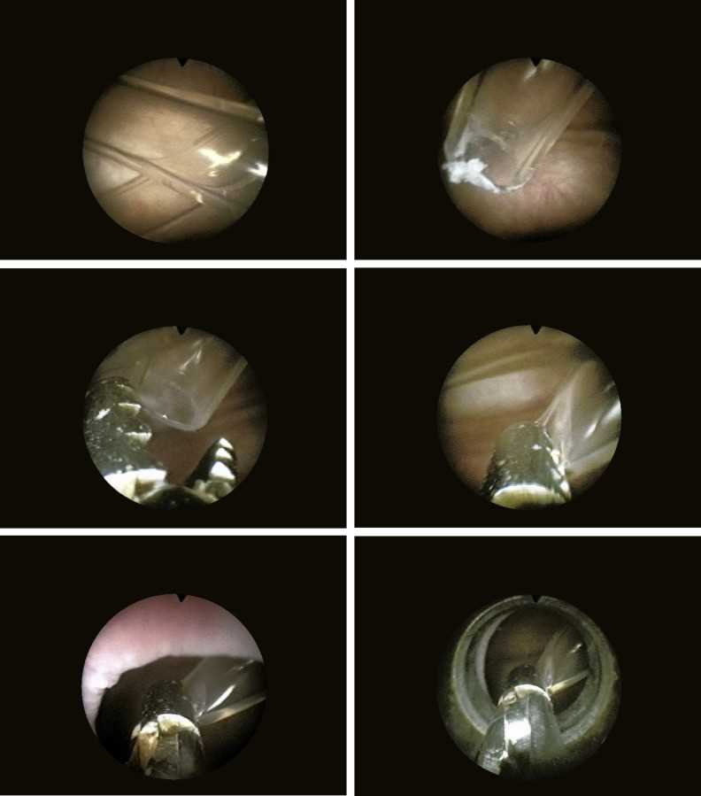
A cystoscope is a medical device designed for visual inspection of the lower urinary tract, particularly the urinary canal. It consists of a long, flexible or rigid tube with a small camera at the tip, allowing healthcare professionals to see real-time images of the affected areas. This tool is commonly employed in procedures where a detailed view is necessary to diagnose conditions such as infections, blockages, or tumors.
By providing a direct visual path into the urinary system, the cystoscope helps doctors detect issues that may not be apparent through external examinations or imaging techniques like X-rays or ultrasound. This procedure, known as cystoscopy, is minimally invasive and often done in a clinic or outpatient setting.
Key Features of a Cystoscope
- Small Camera: The camera at the tip of the device provides high-resolution images, allowing doctors to inspect the urinary passage in detail.
- Flexible or Rigid Design: Depending on the specific examination, the cystoscope may be flexible for easier maneuverability or rigid for a more stable and precise view.
- Light Source: A built-in light at the end of the tool illuminates the area, ensuring clear visibility during the procedure.
- Channels for Tools: Many cystoscopes feature additional channels through which medical instruments can be inserted for tissue sampling or treatment.
Applications of Cystoscopy
- Diagnosis: Cystoscopes are frequently used to diagnose urinary tract infections, bladder cancer, kidney stones, and other related conditions.
- Biopsy: During the procedure, tissue samples can be collected for further testing, aiding in the diagnosis of conditions such as cancer.
- Treatment: In addition to diagnosis, cystoscopes can be used to remove small tumors, treat stones, or perform other minor surgical interventions.
This tool plays a critical role in both diagnosing and treating urinary tract conditions, offering a clear, non-invasive method for assessing the health of the urinary system.
How Cystoscopy Works
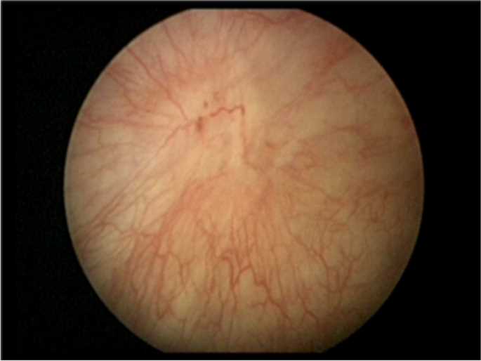
Cystoscopy is a medical procedure that allows doctors to inspect the urinary tract using a specialized tool with a small camera and light. The device is inserted through the urethra and carefully guided into the urinary system. This process provides clear images of the affected area, helping the physician diagnose and treat various conditions. The procedure is minimally invasive and typically performed in an outpatient setting, offering a quick and efficient method of assessment.
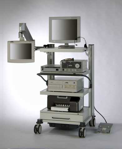
During the procedure, the patient may be given a local anesthetic to minimize discomfort. The flexible or rigid device is then inserted, and the camera sends real-time images to a monitor. The doctor can assess the walls of the urinary canal and surrounding tissues for any abnormalities, such as infections, blockages, or growths. In some cases, small instruments can be passed through the device for tissue biopsy or to remove stones and tumors.
In most cases, cystoscopy is performed under local anesthesia, but in certain situations, sedation or general anesthesia may be used. The procedure typically lasts between 5 and 30 minutes, depending on the complexity of the condition being treated or diagnosed.
Types of Tools for Urinary Tract Inspection
When assessing the health of the urinary system, medical professionals rely on various devices, each designed for specific diagnostic needs. These tools range from flexible models that allow easy navigation through the urinary passage to more rigid types that provide a high degree of precision. The choice of device depends on the condition being diagnosed, the area of the system that needs to be inspected, and the patient’s individual circumstances.
Below are some of the most commonly used devices for inspecting the urinary tract, each with unique features that make it suitable for different diagnostic or therapeutic purposes.
| Device Type | Function | Advantages | Limitations |
|---|---|---|---|
| Flexible Scope | Allows for inspection of the urinary tract with a flexible, easily maneuverable tube. | Minimally invasive, less discomfort for the patient, versatile. | May provide less detailed images than rigid scopes. |
| Rigid Scope | Provides a clearer, more stable view of the urinary system, often used for more complex cases. | High image resolution, ideal for detailed diagnostics. | Can be more uncomfortable, requires local anesthesia. |
| Video Cystoscope | Equipped with a small camera that transmits images to a monitor for real-time visualization. | Allows for clear, high-definition viewing, helpful for documentation. | Higher cost, may require more specialized training to use effectively. |
| Rigid Endoscope | Typically used for larger or more complex examinations, providing detailed imaging of the area. | Great for larger procedures and when precision is required. | Can be uncomfortable and requires a skilled operator. |
| Thin Endoscope | Smaller, more delicate tool, often used for specific, narrow sections of the urinary tract. | Minimally invasive, ideal for more delicate or localized conditions. | May not provide as high-quality imaging as larger models. |
Importance of Urinary Tract Inspection

Assessing the health of the urinary system is crucial for diagnosing a wide range of conditions, from infections to more serious issues like tumors or blockages. A direct view of the urinary canal and surrounding areas allows doctors to accurately identify abnormalities that may not be detectable through standard imaging techniques or physical exams. By visually inspecting these areas, physicians can gain essential insights into the underlying causes of symptoms such as pain, blood in the urine, or difficulty urinating.
Early detection of issues like infections, stones, or tumors can significantly improve treatment outcomes and prevent further complications. Additionally, this type of inspection is vital for monitoring chronic conditions or post-surgical recovery, ensuring that patients receive the appropriate care at every stage of their treatment journey.
Early Diagnosis of Serious Conditions
Immediate access to detailed visuals allows doctors to diagnose serious conditions like bladder cancer, urinary tract infections, and kidney stones at an early stage, leading to faster and more effective treatment. Early intervention can make a substantial difference in patient recovery and quality of life.
Minimally Invasive and Accurate
Direct visual inspection through modern tools allows for a less invasive alternative to traditional diagnostic procedures, minimizing discomfort for the patient while still providing a clear and accurate diagnosis. This method reduces the need for more invasive surgeries, offering a quicker recovery time and less risk for complications.
When is Urinary Tract Inspection Necessary?
There are several situations where a detailed visual assessment of the urinary system becomes essential. These examinations help in identifying underlying issues that may not be detected by other methods, such as blood tests or X-rays. They are particularly useful when symptoms are persistent, unexplained, or severe, and a more thorough investigation is required to reach an accurate diagnosis. Detecting problems early can lead to better outcomes and allow for timely treatment.
Physicians often recommend this type of evaluation if a patient experiences symptoms such as blood in the urine, frequent or painful urination, or unexplained lower abdominal pain. It is also necessary for patients with a history of urinary tract infections or kidney stones, as this procedure can help identify recurring issues or complications.
Common Symptoms That Indicate the Need for Inspection
- Blood in the Urine: Any visible or microscopic blood in the urine can be a sign of infection, stones, or even cancer, necessitating further investigation.
- Painful Urination: If pain or discomfort persists during urination, it may indicate an infection, inflammation, or structural problem in the urinary tract.
- Frequent Urination: Unexplained frequent urges to urinate, especially if accompanied by pain, may require a closer look to rule out underlying issues.
When Monitoring Pre-existing Conditions
For individuals with a history of chronic urinary tract conditions or after undergoing treatment for bladder-related diseases, regular visual inspections are necessary. These assessments ensure that there are no recurrent issues or complications, such as the formation of new stones or the return of infections.
Cystoscope vs. Other Diagnostic Methods
When diagnosing issues related to the urinary system, healthcare professionals have several tools at their disposal. Each diagnostic approach offers distinct advantages depending on the nature of the symptoms and the suspected condition. Among these methods, cystoscopy provides a direct and detailed visual inspection, which can be a crucial advantage when more general tests do not yield enough information. However, other techniques like imaging scans, urine tests, and physical examinations also play important roles in diagnosis.
Understanding the differences between these diagnostic options helps physicians determine the most appropriate approach for each patient. While cystoscopy provides real-time visuals and the ability to perform minor treatments, it may not always be necessary or the most suitable for every case.
Comparison with Imaging Techniques
- X-rays and Ultrasound: These non-invasive imaging methods can help detect stones, tumors, and blockages but do not offer the detailed, real-time views that cystoscopy provides. They are often used as preliminary tools before proceeding with a more invasive procedure.
- CT Scans and MRI: While highly detailed, these methods are often used to detect larger issues or abnormalities. They can offer 3D imaging, but they do not allow for direct intervention or sampling, unlike cystoscopy.
Comparison with Urine Tests and Blood Work
- Urine Tests: Commonly used to detect infections or abnormalities in the urine, these tests are useful for diagnosing urinary tract infections (UTIs) or kidney problems. However, they lack the ability to show structural issues in the urinary system, which cystoscopy can easily reveal.
- Blood Work: Blood tests can provide valuable information about kidney function and the presence of infections or systemic conditions, but they do not directly assess the condition of the urinary tract itself.
In summary, while cystoscopy offers the advantage of direct visual access and allows for precise diagnosis and treatment in real time, other diagnostic methods can complement it by providing initial insights or detailed images when needed. The choice of technique depends on the symptoms, suspected condition, and the healthcare provider’s evaluation of the situation.
Preparation for Urinary Tract Inspection
Preparing for a urinary tract inspection is an essential step to ensure that the procedure goes smoothly and provides accurate results. Proper preparation helps minimize discomfort and allows healthcare providers to gather the necessary information for diagnosis or treatment. The steps involved in getting ready for the procedure can vary based on the method being used and the individual patient’s condition, but there are common guidelines that most patients will follow.
Before the procedure, doctors typically explain what will happen during the process, answer any questions, and provide instructions on how to prepare. This may include fasting for a certain period or adjusting medications that could interfere with the examination. It’s also important to inform the healthcare provider of any allergies, existing medical conditions, or previous urinary tract issues that might affect the procedure.
Dietary and Fluid Restrictions
- Hydration: In many cases, patients may be asked to drink plenty of fluids before the procedure. This ensures that the urinary system is full, making it easier for the doctor to see the areas of interest.
- Fasting: Depending on the type of sedation used during the procedure, fasting for a few hours prior may be necessary. It’s important to follow the doctor’s specific instructions to avoid complications.
Medications and Pre-Procedure Care
- Medication Adjustments: Some medications, such as blood thinners or diuretics, may need to be temporarily adjusted before the procedure. This reduces the risk of bleeding or complications during the examination.
- Antibiotics: If there is a risk of infection, your doctor may prescribe antibiotics before the procedure to reduce the chances of complications.
Following these preparation steps can help ensure that the examination is as effective and comfortable as possible, leading to accurate results and an overall smoother experience for the patient.
Procedure and Techniques of Cystoscopy
Cystoscopy is a minimally invasive procedure that allows doctors to directly view the urinary system for diagnosis and treatment. This procedure typically involves inserting a small, flexible tube through the urethra, providing clear visuals of the urinary canal and surrounding tissues. The process helps in identifying conditions like infections, tumors, blockages, or stones, and can also be used for minor treatments, such as removing small growths or taking tissue samples for biopsy.
The procedure is generally performed in an outpatient setting, and patients are usually given local anesthesia to numb the area. In some cases, sedation or general anesthesia may be used depending on the complexity of the examination and the patient’s comfort level. The doctor will guide the tube carefully into the urinary system while monitoring the real-time images on a screen.
Step-by-Step Process
- Preparation: The patient is asked to lie down, and a local anesthetic or sedative is applied to ensure comfort. A sterile solution is often used to clean the area before insertion.
- Insertion: The doctor gently inserts the flexible tube through the urethra. As the tube moves through the urinary canal, the camera sends images to a monitor for clear visualization.
- Inspection: Once in place, the doctor carefully examines the walls and lining of the area for any signs of infection, blockages, or abnormal growths. If necessary, small tools may be passed through the tube for further intervention, such as taking a biopsy or removing stones.
- Completion: After the examination, the tube is removed, and the procedure is typically concluded within 5 to 30 minutes, depending on the specifics of the case.
Post-Procedure Care
After the procedure, patients are usually allowed to go home the same day. They may experience mild discomfort, such as a sensation of urgency or mild pain during urination. These symptoms are temporary and usually subside within a few days. Patients are advised to drink plenty of fluids and follow any additional instructions provided by the doctor for a smooth recovery.
Common Conditions Diagnosed with Cystoscopy
Cystoscopy is an essential diagnostic tool for identifying a wide range of conditions that affect the lower urinary tract. It allows healthcare providers to directly view the urinary canal, offering a clear perspective on various abnormalities. This procedure can be used to detect infections, structural issues, tumors, and other conditions that might not be easily identified through other diagnostic methods. By providing real-time visuals, cystoscopy helps physicians make accurate diagnoses and plan effective treatment strategies.
Some of the most common conditions diagnosed using this procedure include urinary tract infections, bladder cancer, kidney stones, and chronic pelvic pain. Early detection of these conditions can significantly improve treatment outcomes, allowing for timely intervention and better management of symptoms.
Urinary Tract Infections (UTIs)
Although UTIs are often diagnosed with urine tests, cystoscopy is sometimes recommended for persistent or recurrent infections that do not respond to typical treatments. The procedure helps identify structural issues or abnormalities in the urinary tract that may be contributing to the infections.
Bladder Cancer
Cystoscopy is one of the most reliable methods for detecting bladder cancer. It allows doctors to visually inspect the walls of the urinary system for tumors or abnormal growths. If suspicious areas are found, a biopsy can be performed during the procedure to confirm the diagnosis and determine the best course of treatment.
Kidney Stones

In cases where kidney stones are suspected but not visible on imaging tests, cystoscopy can provide a direct view of the urinary tract to identify any obstructions caused by stones. It can also help in the removal of smaller stones or guide further treatment for larger ones.
Chronic Pelvic Pain and Bladder Disorders
For patients experiencing unexplained chronic pelvic pain, cystoscopy can help identify conditions such as interstitial cystitis, which causes bladder inflammation. The procedure can also reveal other disorders that may be contributing to symptoms like pain or discomfort during urination.
Overall, cystoscopy is a valuable procedure that helps diagnose a variety of conditions affecting the urinary system, offering essential insights that contribute to effective patient care and treatment.
Risks and Complications of Urinary Tract Inspection
While a urinary tract inspection is generally a safe and minimally invasive procedure, there are some risks and potential complications that patients should be aware of. As with any medical procedure, the risk of side effects or adverse reactions depends on various factors, including the patient’s overall health, the complexity of the examination, and the skill of the healthcare provider. It is essential for patients to understand these risks before undergoing the procedure so they can make informed decisions about their care.
Most people experience only minor discomfort during and after the procedure, but in some cases, more serious complications can arise. These may include infections, bleeding, or injury to surrounding tissues. While such complications are rare, it is important to monitor for any signs that may indicate a problem.
Common Risks
- Infection: The most common risk is infection, which can occur when bacteria are introduced into the urinary tract during the procedure. Patients may be prescribed antibiotics to prevent or treat infections if necessary.
- Bleeding: Mild bleeding or spotting is a normal side effect in some cases, but excessive or prolonged bleeding may indicate a more serious issue that requires medical attention.
- Discomfort: Some patients may experience temporary discomfort or a sensation of urgency during urination after the procedure. These symptoms usually subside within a few hours to a few days.
Less Common but Serious Complications
- Urinary Retention: In rare cases, patients may have difficulty urinating after the procedure. This may require further intervention, such as catheterization, to relieve the bladder.
- Tissue Injury: Although uncommon, there is a small risk of injury to the lining of the urinary tract or surrounding organs, particularly if the procedure is more complex or if the patient has certain underlying conditions.
- Allergic Reactions: Some patients may have an allergic reaction to the anesthetic or contrast dye used during the procedure. Symptoms could include itching, swelling, or difficulty breathing, requiring immediate medical attention.
While the risks associated with urinary tract inspections are generally low, it is important for patients to communicate openly with their healthcare provider about any pre-existing conditions, allergies, or concerns. This will help ensure the procedure is as safe and effective as possible. If any unusual symptoms arise after the procedure, it is important to seek medical advice promptly.
Recovery After Urinary Tract Inspection
After undergoing a urinary tract inspection, most patients can expect a relatively short recovery period, with minimal downtime. However, it’s important to follow post-procedure care instructions carefully to ensure a smooth recovery and avoid potential complications. The process typically involves managing mild discomfort, staying hydrated, and monitoring for any unusual symptoms. Recovery varies depending on individual health, the complexity of the procedure, and whether any additional treatments were performed.
Patients are usually able to return to their normal activities within a day or two, but some care is required to avoid irritation or infections. The following guidelines help manage recovery and promote healing after the procedure.
Post-Procedure Care Tips
- Hydration: Drink plenty of water after the procedure. This helps flush out any remaining particles or fluids introduced during the inspection, and it can also reduce any discomfort during urination.
- Avoid Strenuous Activity: It’s advisable to rest and avoid heavy physical activity for 24-48 hours after the procedure. This allows the body time to recover and reduces the risk of complications.
- Manage Discomfort: Some discomfort, such as a mild burning sensation while urinating, is common after the procedure. This should subside within a few days. Over-the-counter pain relievers, like acetaminophen, can be used as recommended by your doctor.
Signs to Watch for During Recovery
- Severe Pain: If you experience intense or worsening pain after the procedure, it may indicate a complication that requires medical attention.
- Heavy Bleeding: While mild spotting is normal, heavy bleeding or blood clots that persist beyond 24 hours should be reported to your healthcare provider.
- Fever: A fever could signal an infection. If your temperature rises above 101°F (38.3°C), contact your doctor.
- Difficulty Urinating: While temporary difficulty urinating can occur, persistent issues or complete inability to urinate should be addressed by your healthcare provider.
By following these recovery guidelines and keeping an eye out for any concerning symptoms, most patients can expect to heal quickly and resume normal activities without complications. If any unusual symptoms persist or worsen, it is essential to contact your healthcare provider for further guidance.
Alternative Methods for Bladder Inspection
While direct visual inspection of the urinary system is a commonly used method for diagnosing various conditions, there are alternative techniques available that can also provide valuable insights. These methods may be preferred in certain situations, depending on the patient’s condition, the complexity of the diagnosis, and any contraindications to more invasive procedures. Each alternative approach offers its own set of benefits, risks, and limitations.
Some methods rely on imaging technology, while others focus on non-invasive techniques to gather information about the urinary tract. These alternatives can be used either as the primary diagnostic tool or in conjunction with other tests to enhance accuracy and reduce discomfort for patients.
Ultrasound Imaging
Ultrasound is one of the most common alternatives for inspecting the urinary system. It uses sound waves to create images of the organs and structures within the abdomen and pelvis, including the kidneys and urinary tract. This method is non-invasive, relatively painless, and does not involve exposure to radiation. Ultrasound is particularly useful for detecting kidney stones, blockages, and structural abnormalities, although it may not provide the same level of detail as direct visual inspection.
CT Scan (Computed Tomography)
A CT scan, or computerized tomography, is another advanced imaging technique that provides detailed cross-sectional images of the body. A CT scan can help identify issues such as tumors, stones, or blockages in the urinary tract. This method is more detailed than ultrasound and can be a valuable tool when a clear visual inspection is required. However, it does involve exposure to radiation, so it may not be the first choice for every patient.
While these alternative methods can offer valuable insights, they may not replace direct visual inspection entirely. In some cases, they can be used to gather preliminary information or to complement other diagnostic procedures, making it easier to determine the next steps for treatment or further investigation.
How to Choose the Right Tool
Selecting the appropriate method for inspecting the urinary system depends on several factors, including the patient’s specific symptoms, medical history, and the goal of the examination. Healthcare providers must assess these aspects to determine which approach will provide the most accurate results while minimizing discomfort and risk to the patient. The right choice can also vary based on whether the goal is a simple diagnosis, treatment of a specific condition, or a more detailed investigation.
Different techniques have distinct advantages and are suited to different situations. For example, some methods are less invasive but may provide less detailed information, while others offer a clearer view but may come with increased risks. Understanding the pros and cons of each option can help both the patient and doctor make an informed decision.
Considerations When Choosing a Method
- Severity of Symptoms: If the patient has severe symptoms or complex issues, a more detailed approach, such as direct visual inspection or advanced imaging, may be necessary.
- Risk Tolerance: Some methods are minimally invasive and carry fewer risks, while others involve a higher degree of risk. The patient’s comfort level and medical history must be taken into account.
- Purpose of the Procedure: The goal of the procedure–whether it’s for diagnosis, monitoring, or treatment–can affect the choice of method. For instance, a simple infection may be diagnosed with a non-invasive method, while a more complex condition might require a more direct approach.
Types of Approaches
- Non-Invasive Imaging: Techniques such as ultrasound and CT scans provide detailed images without the need for insertion into the body. These methods are ideal for patients who need a less invasive approach or when a preliminary overview is needed.
- Direct Visual Inspection: For more precise diagnosis or when a treatment is required alongside diagnosis, a direct inspection might be the best choice. This method is commonly used for detecting tumors, stones, and other abnormalities.
The choice between these methods ultimately depends on the individual case and the healthcare provider’s recommendation. Consulting with a doctor about the benefits, risks, and expected outcomes of each approach can help ensure the best choice for the patient’s needs.
Recent Advances in Bladder Examination Technology
In recent years, significant strides have been made in the technology used to inspect the urinary system. These advances have led to more accurate, less invasive, and faster diagnostic procedures, improving the overall patient experience. With the development of new tools and techniques, healthcare providers can now detect a broader range of conditions with greater precision and fewer risks.
Innovations in imaging technology, such as high-definition endoscopic systems, 3D imaging, and enhanced imaging techniques, are transforming how conditions affecting the urinary tract are diagnosed and treated. These technologies allow for real-time visualization with better resolution, enabling doctors to detect subtle abnormalities that might have previously gone unnoticed.
High-Definition Endoscopy
One of the most notable advancements is the development of high-definition endoscopes. These devices provide a clearer, more detailed view of the urinary system, allowing doctors to identify small tumors, stones, or other abnormalities with greater precision. The high-resolution images help to improve diagnostic accuracy and assist in planning treatment strategies. Additionally, many of these devices are designed to be more flexible and less invasive, leading to a more comfortable experience for patients.
3D Imaging and Virtual Cystoscopy
Another groundbreaking advancement is the use of 3D imaging and virtual cystoscopy. These technologies allow for the creation of three-dimensional reconstructions of the urinary tract based on CT or MRI scans. Virtual cystoscopy offers many of the benefits of traditional methods without the need for physical insertion of a device. It can help detect conditions like tumors, stones, and structural abnormalities with a high degree of accuracy, while reducing patient discomfort and recovery time.
These recent technological advancements are reshaping the field of urinary diagnostics. As they continue to evolve, we can expect even more efficient, non-invasive methods that prioritize patient comfort while ensuring accurate and timely diagnoses.
Patient Experience During Urinary Tract Inspection
Undergoing a urinary system inspection can be an uncomfortable experience for some patients, but advancements in technology have made these procedures increasingly more tolerable. Whether the process is diagnostic or therapeutic, the overall experience depends largely on the method used, the patient’s individual health, and how well the procedure is explained beforehand. Many people feel nervous or anxious about the process, especially when it involves unfamiliar equipment or techniques.
While the goal is always to make the experience as smooth and stress-free as possible, it’s important for patients to understand what to expect during the procedure. Clear communication with the healthcare provider is key to alleviating anxiety and ensuring that patients feel prepared and informed. For most individuals, the discomfort is temporary, and the benefits of accurate diagnosis far outweigh any momentary unease.
What to Expect During the Procedure
- Preparation: Before the procedure, patients may be asked to drink fluids to ensure the area of interest is adequately visible. They may also be advised to avoid urinating for a certain period of time to ensure an optimal examination.
- Discomfort: Mild discomfort is common, particularly when a small device is inserted into the urinary tract. Patients may feel a sensation of pressure or a slight burning, but these feelings typically subside once the procedure is over.
- Duration: Most examinations take only a few minutes, although this can vary depending on the complexity of the situation. Patients are usually able to go home the same day, with no extended recovery time required.
After the Procedure
- Temporary Symptoms: It’s normal to experience slight discomfort, a burning sensation when urinating, or mild bleeding after the inspection. These symptoms usually resolve within a short period.
- Follow-Up Care: In some cases, patients may need a follow-up appointment to discuss the results of the inspection and any subsequent treatments. This is a good time to ask any remaining questions or express concerns.
By understanding the procedure and communicating openly with their healthcare provider, patients can feel more at ease and better prepared for the experience. The goal is always to minimize discomfort while ensuring that any underlying issues are thoroughly examined and appropriately addressed.
Future Trends in Bladder Diagnostics
The future of urinary system diagnostics is poised for significant advancements, driven by technological innovation and a deeper understanding of patient needs. With the rise of more accurate, non-invasive methods, the focus is shifting toward improving both the precision of diagnoses and the overall patient experience. These advancements will not only help detect conditions earlier but also offer quicker, safer, and more comfortable procedures for individuals requiring examination.
Emerging technologies such as artificial intelligence (AI), machine learning, and advanced imaging techniques are expected to revolutionize how medical professionals detect and monitor various conditions affecting the urinary tract. These tools can enhance diagnostic accuracy, reduce the need for invasive procedures, and even help predict future complications, allowing for more proactive care.
Innovations on the Horizon

Several key trends are shaping the future of diagnostic procedures for urinary system health:
- Artificial Intelligence (AI): AI algorithms are being integrated into diagnostic imaging tools to help identify abnormalities such as tumors or stones with greater accuracy. Machine learning models can analyze vast amounts of data to detect patterns that human clinicians may miss, thus improving early detection rates.
- Non-Invasive Testing: As patient comfort remains a primary concern, the development of more non-invasive diagnostic techniques is on the rise. Advances in imaging technology, such as high-resolution MRI and ultrasound, allow doctors to get detailed views of the urinary tract without the need for physical insertion of devices.
- Personalized Diagnostics: Genetic testing and biomarker analysis are making it possible to tailor diagnostic approaches to individual patients. This could enable doctors to predict the risk of developing certain conditions and select the most appropriate diagnostic method based on the patient’s unique characteristics.
Technology-Driven Efficiency
The integration of these emerging technologies will help streamline diagnostic processes, reducing waiting times and improving the efficiency of healthcare systems. Enhanced data collection and analysis capabilities will also provide physicians with more comprehensive insights into a patient’s condition, leading to better-informed decisions and more effective treatments.
| Technology | Benefits | Challenges |
|---|---|---|
| Artificial Intelligence | Improved accuracy, early detection, pattern recognition | High costs, need for data training, reliance on algorithms |
| Non-Invasive Imaging | Less discomfort, quicker procedures, no need for insertion | Possible limitations in visibility for complex conditions |
| Personalized Diagnostics | Tailored care, predictive analytics | Expensive, accessibility challenges, not widely available |
As these technologies continue to develop, we can expect to see even more advancements that improve the diagnostic landscape, making urinary system assessments faster, safer, and more precise for patients and healthcare providers alike. The future of diagnostics promises a more patient-centered, accurate, and efficient approach to care.