
Medical imaging is an essential tool in diagnosing a wide range of health conditions. By using advanced technology, healthcare providers can gain insights into the internal structures of the body, aiding in the detection of various issues that might not be visible through routine physical examinations. These methods play a critical role in determining the cause of symptoms and guiding treatment plans.
When preparing for such a procedure, it’s common for individuals to have numerous inquiries regarding what to expect. From understanding the purpose of the test to learning how to interpret the results, knowing the right information beforehand can make the process less intimidating and more manageable. This section addresses some of the most frequently asked inquiries about diagnostic imaging techniques, providing clear explanations to help clarify any doubts.
Understanding Diagnostic Imaging Procedure
In the field of healthcare, certain procedures are designed to provide detailed images of the body’s internal structures. These techniques help specialists observe organs and tissues, aiding in the identification of potential health issues that may not be visible through routine physical assessments. Such procedures are particularly valuable in diagnosing conditions early, allowing for more effective treatment options.
To gain a comprehensive understanding of this type of imaging, it’s important to know the key components and benefits it offers. This approach is non-invasive, safe, and provides real-time visual data, which enhances diagnostic accuracy. Whether used for monitoring a chronic condition or identifying new health concerns, this method remains an integral part of modern medical diagnostics.
Key advantages of this type of procedure include:
- Non-invasive approach that does not require surgery or incisions.
- Ability to visualize internal organs and tissues in real time.
- Minimal discomfort for the patient during the process.
- Useful for diagnosing a wide range of conditions.
Specialists rely on the results from these procedures to form accurate diagnoses and recommend appropriate treatments. By using sound waves or other methods to capture images, doctors are able to examine various parts of the body in great detail, making it easier to detect abnormalities such as tumors, cysts, or inflammation. This ensures patients receive timely interventions when necessary.
What is Diagnostic Imaging Procedure
This medical procedure uses advanced technology to capture detailed images of the body’s internal structures, helping healthcare providers assess the condition of various organs and tissues. By employing sound waves or other methods, this technique allows specialists to observe areas of the body that are difficult to examine through traditional means. The images produced can help diagnose a range of health conditions, from minor concerns to serious diseases.
One of the primary purposes of this technique is to offer a non-invasive and painless way to gather important data about a patient’s health. By using external sensors or devices, healthcare providers can get a closer look at areas such as blood vessels, muscles, and organs, without the need for surgery or other invasive methods.
Key characteristics of this procedure include:
| Feature | Description |
|---|---|
| Non-invasive | No need for incisions or injections. |
| Real-time Imaging | Produces live visual data for immediate analysis. |
| Minimal Risk | Generally considered safe, with no long-term side effects. |
| Wide Range of Uses | Helpful in diagnosing various conditions, including inflammation, tumors, and blockages. |
These procedures are crucial in helping doctors determine the presence of conditions that may otherwise remain undetected until they become more serious. With its ability to provide clear and accurate images, it has become an indispensable tool in modern healthcare.
Common Reasons for Diagnostic Imaging
This type of diagnostic procedure is commonly used to assess a variety of medical conditions. It provides healthcare professionals with critical visual information to evaluate the health of internal organs and tissues. The non-invasive nature of this method makes it a preferred choice for monitoring, diagnosing, and guiding treatment for a wide range of ailments.
Healthcare providers often recommend this procedure for patients experiencing unexplained symptoms or as part of routine screenings. It can be instrumental in detecting early-stage conditions that may not yet show outward signs or symptoms, allowing for timely intervention and treatment.
Some common reasons for utilizing this diagnostic tool include:
| Reason | Condition or Concern |
|---|---|
| Abdominal Pain | Helps identify issues such as kidney stones, gallbladder disease, or liver problems. |
| Heart Monitoring | Used to examine heart function, blood flow, and potential blockages. |
| Pregnancy Monitoring | Ensures proper fetal development and checks for complications during pregnancy. |
| Musculoskeletal Issues | Evaluates joints, muscles, and soft tissues to detect strains, tears, or injuries. |
| Blood Flow Assessment | Used to check for potential blockages or abnormalities in blood vessels. |
This imaging procedure plays a crucial role in preventing complications and facilitating prompt medical attention when necessary. By providing real-time insights into the body’s internal condition, it enables doctors to make informed decisions regarding diagnosis and treatment plans.
Preparing for Diagnostic Imaging Procedure
Before undergoing a diagnostic imaging procedure, it is important to prepare properly to ensure accurate results and a smooth experience. This preparation may vary depending on the type of procedure and the area of the body being examined. Clear instructions will typically be provided by your healthcare provider, but understanding the general steps involved can help alleviate any concerns.
For many individuals, the preparation involves simple steps such as wearing loose clothing, avoiding certain foods, or refraining from drinking liquids for a specified period of time before the procedure. It is important to follow these guidelines carefully to help the imaging equipment function effectively and to ensure clear, reliable results.
Common preparation steps include:
- Wearing comfortable, loose-fitting clothing to allow easy access to the area being examined.
- Avoiding heavy meals or drinking certain liquids before the procedure, particularly if it involves the abdomen.
- Informing your healthcare provider about any medications or health conditions that might affect the procedure.
- Arriving early to complete any necessary paperwork and to ask any last-minute questions.
- Following any specific instructions related to fasting or hydration depending on the type of examination.
By preparing in advance and following your doctor’s recommendations, you can help ensure the procedure goes smoothly and that the images captured are of the highest quality. This allows healthcare providers to make the most accurate assessments based on the results.
What Happens During Diagnostic Imaging

During a diagnostic imaging procedure, the process is designed to be non-invasive and relatively quick. It involves using specialized equipment to capture detailed images of the body’s internal structures. While the specifics may vary based on the area being examined, the procedure generally follows a consistent sequence of events to ensure accurate results and patient comfort.
Before starting, a healthcare professional will typically explain the procedure, addressing any last-minute questions or concerns. The patient is then positioned comfortably, and the technician will apply a gel or other substance to the area being examined to facilitate better imaging. The procedure itself is generally painless, with the primary focus on capturing clear visual data.
Here is an overview of what typically happens:
- Positioning: The patient will be asked to lie down or sit in a specific position based on the area being examined.
- Gel Application: A special gel is applied to the skin to help transmit sound waves or other signals for better image clarity.
- Imaging Process: A handheld device, often referred to as a sensor or probe, is moved over the skin to capture the necessary images.
- Duration: The procedure usually lasts between 15 to 45 minutes, depending on the complexity and area of examination.
- Monitoring: Throughout the process, the technician will monitor the images in real time and may adjust the equipment to ensure optimal results.
Once the procedure is complete, the gel is wiped off, and the patient can resume normal activities. Any further instructions or follow-up appointments will be provided by the healthcare team based on the results.
Key Benefits of Diagnostic Imaging
Diagnostic imaging plays a crucial role in modern healthcare by providing detailed visual information about the internal structures of the body. The benefits of this non-invasive procedure extend far beyond just diagnosis. It allows for accurate assessments, early detection of conditions, and continuous monitoring of patients’ health, all with minimal discomfort or risk. This method is vital for creating effective treatment plans and improving patient outcomes.
Non-Invasive and Safe
One of the primary advantages of this technique is that it is non-invasive, meaning there is no need for surgery or other intrusive methods to gather critical information. This reduces the risks associated with more invasive procedures and allows for quicker recovery times. Additionally, this imaging method is considered safe, with minimal side effects or long-term health concerns for most patients.
Real-Time Imaging for Accurate Diagnosis
This diagnostic method provides real-time images, allowing healthcare providers to assess the condition of organs and tissues immediately. This dynamic, live feedback enables doctors to make quicker decisions and identify problems early, which is essential for effective treatment. Real-time imaging also facilitates better communication between patients and doctors, as both can view the results together during the procedure.
Other significant benefits include:
- Clear visualization of internal structures, such as muscles, organs, and blood vessels.
- Helpful in monitoring the progress of existing conditions and evaluating the effectiveness of treatments.
- Minimal discomfort, with most patients reporting no pain during the procedure.
- Versatility in diagnosing a wide range of health conditions, from cardiovascular issues to musculoskeletal injuries.
With these key benefits, this imaging technique continues to be an essential tool in healthcare, supporting doctors in delivering accurate diagnoses and timely treatments.
How to Interpret Diagnostic Imaging Results
Interpreting the results from a diagnostic imaging procedure requires both expertise and a keen understanding of how the images reflect the internal structures of the body. The images captured during the procedure provide valuable insights that can indicate normal health conditions or highlight areas of concern. Healthcare providers analyze these results carefully to determine whether further tests or treatments are necessary.
The interpretation process involves reviewing the images and identifying any abnormalities or deviations from expected patterns. In some cases, the images may reveal clear signs of issues such as blockages, cysts, or tissue damage, while in other instances, further tests may be required to confirm any findings. It is important for healthcare professionals to consider the patient’s medical history and symptoms when interpreting the results to ensure an accurate diagnosis.
Key steps in interpreting the results include:
- Assessing Image Clarity: The first step is to ensure that the images are clear and detailed, which helps in accurate analysis. Any blurring or distortion may require a repeat of the procedure.
- Identifying Abnormalities: Specialists look for irregularities such as fluid buildup, inflammation, or masses that could signal a health concern.
- Comparing with Norms: The healthcare provider compares the images to typical reference images to identify what is normal versus what could be considered an anomaly.
- Contextualizing with Symptoms: The results are then interpreted in conjunction with the patient’s reported symptoms and medical history to form a complete picture of the situation.
While interpreting these results can be complex, skilled professionals use their knowledge and experience to guide decisions about next steps, ensuring that patients receive the appropriate care based on the findings.
What to Expect After Diagnostic Imaging
Once a diagnostic imaging procedure is complete, the next steps typically involve a brief recovery period, followed by the interpretation of the images by a healthcare professional. Most patients can resume their daily activities immediately, as the procedure itself is non-invasive and causes minimal discomfort. However, the next phase–waiting for the results–may vary depending on the situation and the urgency of the findings.
After the procedure, healthcare providers will review the images and, if necessary, discuss any follow-up actions with the patient. In some cases, additional tests or procedures may be recommended to gather more detailed information. The timeframe for receiving results can differ, with some cases requiring immediate evaluation, while others may take a few days for analysis.
Immediate Care and Aftercare
For most patients, there is no special aftercare required. However, if the procedure was used to assess specific conditions, your healthcare provider may offer guidance on any temporary lifestyle changes or precautions. These instructions will depend on the area of the body examined and the health concerns being addressed. It is important to follow these recommendations to support your recovery and ensure that no further complications arise.
Understanding the Results
The results of the procedure are typically shared with you in a follow-up appointment or via your healthcare provider. They will explain what the images reveal and discuss whether further steps, such as additional tests or treatment plans, are necessary. If there are concerns or abnormal findings, your doctor will outline the next steps in addressing those issues.
While waiting for the results, it’s important to remain in communication with your healthcare provider to address any questions or concerns that may arise. The professional interpretation of the images will be critical in determining the appropriate course of action for your health.
Who Should Get a Diagnostic Imaging Procedure
Diagnostic imaging is a valuable tool for individuals experiencing health concerns or for those needing routine assessments to monitor their well-being. It can be particularly helpful for diagnosing a wide range of conditions, from soft tissue injuries to chronic diseases. The decision to undergo this type of procedure is typically based on specific symptoms, medical history, and the guidance of a healthcare professional.
While anyone can potentially benefit from this diagnostic technique, certain groups of people may be more likely to require it. In particular, individuals with unexplained pain, ongoing medical issues, or those at higher risk for specific health conditions may be advised to undergo an imaging procedure to gain more clarity about their condition.
Common Reasons for Referral

There are several scenarios in which a healthcare provider may recommend a diagnostic procedure:
- Persistent Pain: Individuals experiencing ongoing or unexplained pain, especially in areas like the abdomen, chest, or joints, may need imaging to identify the underlying cause.
- Chronic Conditions: People with long-term health conditions such as heart disease, arthritis, or diabetes may be monitored with imaging to assess the progress of their condition.
- Injuries: Individuals who have sustained an injury, particularly a sprain, fracture, or muscle tear, may require imaging to evaluate the extent of the damage.
- Abnormal Symptoms: Those experiencing symptoms such as swelling, abnormal growths, or unexplained fatigue may undergo this procedure to rule out serious underlying causes.
Who May Benefit the Most
People who are at higher risk for certain health issues may also be more likely to benefit from regular diagnostic screenings:
- Older Adults: As people age, the risk for conditions like cardiovascular disease, arthritis, and certain cancers increases, making regular imaging a helpful tool for early detection.
- Individuals with a Family History: Those with a family history of specific diseases may be advised to undergo imaging more frequently to monitor for early signs of these conditions.
- High-Risk Populations: People with lifestyle factors such as smoking, high blood pressure, or a sedentary lifestyle may be encouraged to have imaging to detect issues such as cardiovascular problems or lung diseases early.
Ultimately, the decision to undergo this procedure should be made in consultation with a healthcare provider, who can assess the individual’s unique health needs and determine whether diagnostic imaging is necessary.
Comparing Diagnostic Imaging with Other Tests

When assessing health conditions, various diagnostic tests are available, each offering unique advantages depending on the situation. Some procedures are more suited for specific concerns, while others provide a broader range of insights. Understanding the differences between these methods helps healthcare providers choose the most effective approach for diagnosing and managing a patient’s condition. This section compares one common imaging technique with other testing options to highlight their respective benefits and limitations.
Each diagnostic tool has its strengths, whether it’s providing detailed internal imagery, monitoring specific body functions, or evaluating physiological responses. The choice of which test to use often depends on the type of information needed and the patient’s medical history.
| Test Type | Purpose | Advantages | Limitations |
|---|---|---|---|
| Imaging Procedure | Provides detailed images of internal structures | Non-invasive, accurate, quick results | Can be costly, may require preparation |
| Blood Test | Analyzes chemical and biological markers in the body | Inexpensive, easy to perform, offers insight into health conditions | Doesn’t provide detailed internal imagery, limited in scope |
| X-ray | Uses radiation to visualize bones and certain tissues | Quick, effective for detecting fractures and certain diseases | Exposure to radiation, limited in detecting soft tissue issues |
| MRI | Uses magnetic fields to create detailed images of organs and tissues | Highly detailed images, no radiation exposure | Expensive, takes longer to complete, not suitable for patients with metal implants |
While the imaging technique discussed here is particularly useful for evaluating certain conditions, other diagnostic tests such as blood tests, MRIs, and X-rays are invaluable for assessing different aspects of health. Each method provides critical data, and in many cases, a combination of tests is used to achieve a comprehensive understanding of a patient’s condition.
Is Diagnostic Imaging Painful or Risky?
When considering any medical procedure, patients often wonder about the level of discomfort or potential risks involved. Diagnostic imaging is generally regarded as a non-invasive and safe method for assessing health conditions, but it’s important to understand whether there are any associated discomforts or hazards. This section explores whether the procedure causes pain and if there are any potential risks to be aware of.
Discomfort During the Procedure
Most people find that the diagnostic process is relatively painless, although some may experience mild discomfort, particularly if the procedure involves applying pressure to a sensitive area of the body. In most cases, patients are able to tolerate the procedure without significant pain. Factors that might affect the level of discomfort include:
- Positioning: Depending on the area being assessed, certain positions may cause temporary discomfort or stiffness.
- Duration: If the procedure takes longer than expected, there may be some minor discomfort from remaining in one position.
- Sensitivity: Areas of the body that are already sore or inflamed might be more sensitive during the procedure.
Potential Risks and Safety
While diagnostic imaging is typically safe, it is important to consider any potential risks associated with certain procedures. The risk of harm depends largely on the type of test being performed and the individual’s health status. Common concerns include:
- Radiation Exposure: Some imaging tests, such as X-rays, involve radiation, which could pose a risk if done excessively. However, modern imaging techniques are designed to minimize exposure.
- Allergic Reactions: In cases where contrast agents are used, there is a very small risk of an allergic reaction. These reactions are rare but can lead to mild or severe symptoms.
- Claustrophobia: Tests like MRIs, which require patients to stay inside a machine for extended periods, may cause discomfort for those with claustrophobia.
Overall, the risks associated with diagnostic imaging are minimal, and the benefits of obtaining critical health information often outweigh any minor discomfort. Before undergoing the procedure, it is always advisable to discuss any concerns with a healthcare provider to ensure the best possible experience.
Understanding the Role of Technicians
In any diagnostic procedure, the role of the professional operating the equipment is crucial to ensuring the process runs smoothly and accurately. Technicians are specially trained to handle the machinery, assist patients, and ensure that the correct procedure is followed. This section highlights the responsibilities and expertise of these professionals and why they are integral to the entire diagnostic process.
Key Responsibilities of Technicians
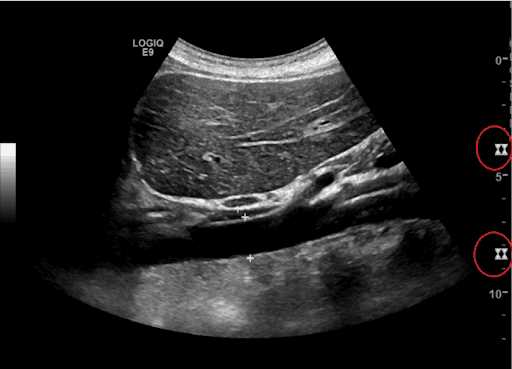
Technicians play a key part in ensuring that diagnostic assessments are conducted effectively. They are responsible for preparing the equipment, assisting patients during the procedure, and ensuring the highest standards of accuracy. Below are some of their main duties:
- Preparation: Technicians ensure that all equipment is set up correctly and ready for use before starting the procedure.
- Patient Assistance: They guide patients through the process, explaining the steps and making sure they are comfortable.
- Monitoring: During the procedure, technicians closely monitor the equipment to ensure it is functioning properly and gather the necessary data.
- Safety: They make sure safety protocols are followed, such as ensuring that no harm comes to the patient during the procedure.
Training and Qualifications
Technicians typically undergo extensive training to ensure they have the skills necessary for performing their duties. Their education includes understanding the technology they use, as well as learning how to interpret basic results. Training programs often involve both classroom instruction and hands-on experience, ensuring that technicians are fully equipped to handle complex equipment and interact with patients in a professional manner.
| Area of Expertise | Importance | Examples |
|---|---|---|
| Technical Knowledge | Ensures that all equipment functions properly and data is captured accurately | Operating diagnostic machinery, troubleshooting issues |
| Patient Interaction | Assists in making the patient feel comfortable and explains the process clearly | Providing reassurance, answering patient questions |
| Safety Protocols | Guarantees the procedure is conducted without harm to the patient | Ensuring proper positioning, minimizing risks during testing |
Technicians are vital to ensuring the success of the diagnostic process. Their expertise and attention to detail ensure that procedures are conducted smoothly, safely, and with minimal discomfort to the patient. Their role helps to make diagnostic assessments both efficient and accurate, which is crucial for proper patient care.
Can SPI Ultrasound Detect Certain Conditions

Diagnostic procedures using specialized equipment can help identify and assess various medical conditions. Certain techniques are particularly effective in revealing issues related to the circulatory system, internal organs, and soft tissue abnormalities. This section explores how such a procedure can be instrumental in detecting various conditions that might otherwise go unnoticed in early stages.
Conditions Identified Through the Procedure
During the procedure, advanced imaging technologies can provide detailed views of internal structures, allowing healthcare professionals to identify specific health issues. Some common conditions that can be detected include:
- Blood Flow Issues: The procedure can reveal blockages or irregularities in blood vessels, helping to diagnose cardiovascular conditions like stenosis or clot formation.
- Organ Abnormalities: Internal organs, such as the liver, kidneys, and heart, can be examined for conditions like cysts, tumors, or signs of disease.
- Infections: Infections that affect organs or tissues, such as abscesses, may be identified through visual imaging.
- Soft Tissue Damage: Conditions such as muscle tears, ligament strains, or other injuries affecting soft tissues can also be detected with the help of this method.
Limitations and Considerations
While the procedure is highly effective in detecting a wide range of conditions, there are certain limitations. Not all issues can be visualized or fully diagnosed through this method. In some cases, additional tests or procedures may be required for a complete diagnosis. Moreover, the clarity of the images can sometimes be affected by factors such as patient body type or the presence of excessive movement during the procedure.
Ultimately, this diagnostic technique is a valuable tool in detecting and monitoring various health conditions, providing early insights that can guide treatment decisions and improve patient outcomes.
How Accurate is SPI Ultrasound Exam
When it comes to diagnostic procedures, accuracy plays a crucial role in ensuring proper treatment and management of medical conditions. The effectiveness of certain imaging techniques is measured by their ability to provide precise and reliable results. In this section, we explore how accurate such procedures are in detecting various conditions, and the factors that can influence their effectiveness.
The reliability of diagnostic procedures depends on several factors, such as the technology used, the skill of the technician, and the patient’s condition. While the procedure is known for its high accuracy in assessing many health conditions, it is important to understand that no method is 100% flawless. Certain challenges can affect the clarity and quality of the results.
Factors Influencing Accuracy
The accuracy of the results can be influenced by the following:
- Technician Expertise: The proficiency of the technician operating the equipment significantly affects the quality of the images obtained. Skilled technicians can position the device more effectively and interpret the results more accurately.
- Patient’s Body Type: Factors like obesity or excessive gas in the abdomen can sometimes interfere with the clarity of the results, making it harder to detect abnormalities.
- Device Quality: The type of technology used during the procedure also determines how precise the results are. Advanced equipment can provide more detailed and clearer images, leading to more accurate diagnoses.
Limitations and Considerations
While the procedure is generally reliable, there are certain limitations to its accuracy. Some conditions may not be easily visible, especially if they are in the early stages or located in difficult-to-reach areas of the body. In such cases, additional tests or imaging techniques may be needed to confirm a diagnosis.
Overall, this diagnostic technique provides a high level of accuracy, especially when performed by an experienced professional. However, as with all medical procedures, it is essential to consider the broader context of a patient’s health and possibly combine results with other diagnostic tools for a comprehensive understanding of their condition.
Cost Considerations for SPI Ultrasound
The financial aspect of medical procedures is a significant concern for many patients. Understanding the potential costs associated with diagnostic imaging plays a crucial role in planning and decision-making. While some procedures may be covered by insurance, others might require out-of-pocket expenses, depending on various factors. In this section, we explore the key elements that contribute to the cost of such assessments.
Several factors influence the overall cost, including the type of facility where the procedure is performed, the geographic location, and whether additional tests are required. It’s important for patients to be aware of the potential expenses involved and consider these when discussing options with their healthcare providers.
Key Factors Affecting Costs
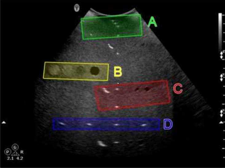
The cost of diagnostic assessments is determined by a variety of factors:
- Facility Type: Hospitals and specialized clinics often charge different rates. Facilities that are more equipped with advanced technology or offer more comprehensive care tend to have higher fees.
- Location: The cost can vary significantly based on the region. Urban areas with more medical centers might have competitive pricing, while rural areas may have fewer options, potentially increasing costs.
- Insurance Coverage: Some insurance plans cover the procedure entirely or partially, while others may require co-pays or deductible payments. Patients should check with their insurance provider to understand their coverage.
- Additional Tests: If further diagnostic procedures are needed to confirm results or investigate additional concerns, the cost can rise accordingly. Patients should inquire about the need for follow-up tests.
Average Costs and Payment Options
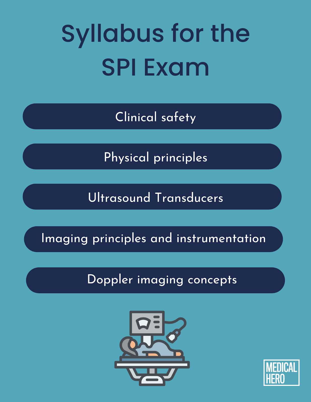
While prices can vary, on average, patients can expect to pay anywhere from a few hundred to several thousand dollars for diagnostic assessments, depending on the factors mentioned above. It’s advisable to request an estimate beforehand from the healthcare provider to ensure transparency.
For those without insurance or who face high deductibles, exploring payment plans or discussing financial assistance with the healthcare provider may help manage costs. Some facilities offer sliding scale fees based on income, which can make these procedures more accessible.
Ultimately, understanding the cost structure and considering financial options ensures that patients can make informed decisions about their healthcare while balancing their budgetary needs.
SPI Ultrasound Exam vs Traditional Methods
In the field of medical diagnostics, advancements in technology have introduced newer methods for assessing and monitoring the body. These modern techniques often provide quicker results and may be less invasive compared to older, traditional methods. In this section, we compare the benefits and limitations of cutting-edge imaging technologies with more conventional diagnostic approaches, highlighting their respective roles in patient care.
Both modern and traditional diagnostic methods serve the same purpose: to provide accurate insights into a patient’s condition. However, the tools and techniques used differ significantly in terms of complexity, procedure time, cost, and overall patient comfort.
Advantages of Modern Techniques
Recent developments in diagnostic procedures have brought several advantages that are particularly beneficial to patients:
- Less Invasive: Many modern technologies, such as imaging techniques, do not require surgical intervention, reducing the risks of infection and recovery time.
- Quick and Painless: These methods typically take less time to complete and do not involve discomfort for the patient, improving the overall experience.
- High Accuracy: Newer technologies often provide higher resolution images and more precise data, allowing for better diagnosis and treatment planning.
- Non-Radiation Options: Many advanced methods, unlike traditional X-rays or CT scans, do not involve exposure to harmful radiation, making them safer for frequent use.
Challenges of Traditional Diagnostic Techniques
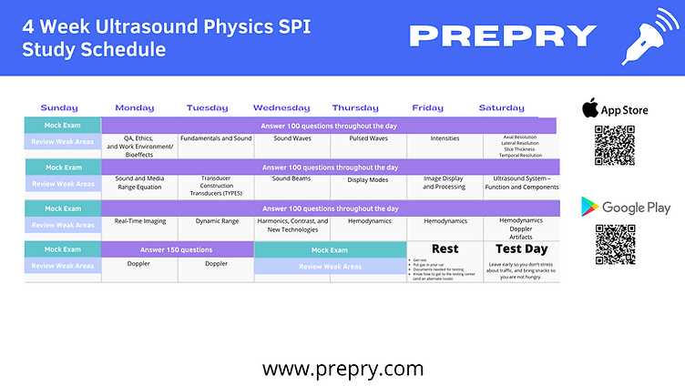
While traditional diagnostic methods, such as X-rays or MRI scans, have been used for decades, they come with certain drawbacks:
- Longer Procedure Times: Older methods can often take more time to complete, leading to extended patient wait times and longer overall appointments.
- Invasiveness: Some traditional methods may require more invasive procedures, such as biopsies or injections, to obtain necessary data, which can cause discomfort and carry higher risks.
- Higher Costs: Many traditional diagnostic techniques, such as CT scans, can be more expensive, both for the patient and the healthcare system.
- Radiation Exposure: Some older methods involve radiation, which may increase the risk of long-term health concerns with repeated exposure.
Ultimately, both modern and traditional diagnostic techniques have their place in healthcare. The choice of method depends on the patient’s condition, the healthcare provider’s judgment, and the desired outcome of the procedure. While advanced methods offer certain advantages in terms of efficiency and comfort, traditional approaches are still invaluable in specific diagnostic scenarios.
FAQ About SPI Ultrasound Procedure
In this section, we address some of the most common inquiries people have regarding the procedure. Understanding the process and what to expect can help reduce any uncertainties and make the experience more comfortable. Below, we answer questions related to preparation, safety, and what happens during the procedure itself.
What is the purpose of this diagnostic procedure?
This technique is primarily used to assess internal organs, blood flow, and other body systems without the need for invasive surgery. It provides detailed images that help doctors evaluate conditions and plan appropriate treatments.
How should I prepare for the procedure?
Preparation depends on the specific area of the body being evaluated. In general, you may be asked to refrain from eating or drinking for a few hours before the procedure. It’s essential to follow any instructions provided by your healthcare provider to ensure accurate results.
Is the procedure painful?
No, this procedure is generally painless. The process involves applying a gel to the skin and using a device to capture images. You may feel slight pressure, but discomfort is minimal. If you experience any pain or discomfort, inform the technician immediately.
Are there any risks associated with this procedure?
This diagnostic tool is non-invasive and considered very safe. Unlike other imaging techniques that use radiation, it poses no risk of exposure to harmful substances. However, if you are pregnant or have any specific health conditions, it’s important to inform your doctor beforehand.
How long does the procedure take?

The procedure usually lasts between 30 minutes to an hour, depending on the area being examined. You may be asked to remain still during the process to ensure clear images are obtained.
When will I get the results?
In most cases, the results are available shortly after the procedure is completed. Your healthcare provider will explain the findings to you and discuss any next steps if further action is needed.
Are there any special aftercare instructions?
There are generally no specific aftercare instructions following the procedure. You can resume normal activities immediately. If you experience any unusual symptoms, such as discomfort or swelling, be sure to contact your healthcare provider.