As medical imaging technology continues to evolve, understanding the process of advanced scanning techniques is essential for both patients and healthcare professionals. These imaging methods play a critical role in diagnosing a wide range of conditions, allowing for accurate assessments and treatment plans.
The purpose of this section is to provide essential insights into the most commonly asked topics related to these diagnostic scans. By familiarizing yourself with the fundamental aspects, you can feel more confident and prepared when navigating through the procedures and their associated concepts.
From safety guidelines to interpretation of results, each detail is crucial in ensuring a smooth experience and accurate outcome. Whether you’re preparing for a medical career or simply looking to gain more knowledge, this guide offers a comprehensive overview to help you succeed.
MRI Exam Questions and Answers
This section provides an in-depth exploration of common topics that often arise in relation to advanced scanning procedures. It covers a range of practical scenarios, from technical specifications to safety measures, all aimed at ensuring a thorough understanding of the diagnostic method. Familiarizing yourself with these key points can enhance both theoretical knowledge and practical skills.
By reviewing typical inquiries and their corresponding solutions, individuals can better prepare for handling the various aspects of these medical techniques. This information is particularly useful for those looking to solidify their grasp on the fundamental principles, whether for academic purposes or practical application in a clinical environment.
Each element of the scanning process, from patient preparation to machine operation, is broken down into digestible sections, helping to clarify complex concepts. Understanding the expected procedures and how to respond in different situations is crucial for achieving accurate results and maintaining safety during the process.
Common MRI Exam Topics You Should Know
Understanding the core concepts related to advanced imaging procedures is crucial for both aspiring professionals and individuals looking to deepen their knowledge. This section focuses on the most important topics that frequently come up during evaluations of these diagnostic techniques. Grasping these concepts will help you navigate the complexities of the process and prepare for practical application.
Key Principles of Imaging Techniques
One of the foundational areas to explore is the basic principles behind how these scans work. Key topics include:
- Magnetic fields and their role in creating images
- The mechanics of how images are generated
- Understanding signal generation and reception
Patient Preparation and Safety Guidelines
Safety is always a top priority, and knowing how to prepare patients is essential. This involves:
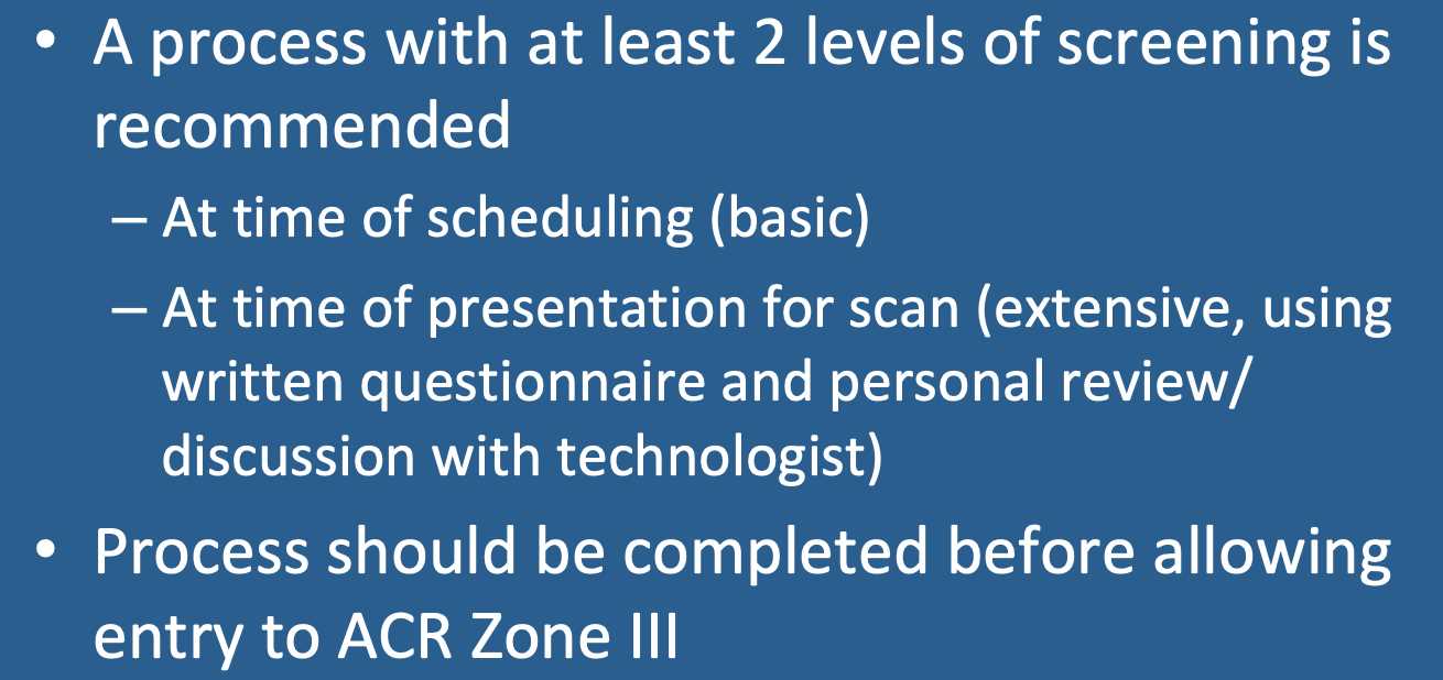
- Pre-procedure screenings to ensure patient safety
- Identifying contraindications (e.g., implants, medical devices)
- Proper positioning and comfort for accurate results
Mastering these topics will give you the foundational knowledge required to succeed in understanding and applying this diagnostic technology. Each of these areas plays a vital role in the success of the procedure and the quality of the resulting images.
How to Prepare for Your MRI Test
Preparing for an advanced diagnostic scan is a crucial step in ensuring the process goes smoothly. Proper preparation not only helps with the accuracy of the results but also reduces anxiety for the patient. Understanding the necessary steps and guidelines can make the entire experience much more comfortable and efficient.
Pre-Procedure Instructions
Before undergoing a diagnostic scan, there are several important things to keep in mind. These preparations ensure that the procedure runs smoothly and that all necessary precautions are taken.
| Preparation Step | Details |
|---|---|
| Health Screening | Inform your healthcare provider about any medical conditions or implants, as some may interfere with the process. |
| Clothing | Wear loose, comfortable clothing without any metal zippers or buttons, as they can affect the scan. |
| Fasting | For certain procedures, you may be asked to avoid eating or drinking for a few hours before the test. |
What to Expect on the Day of the Procedure
On the day of the scan, it’s important to arrive early to complete any necessary paperwork. You will be given instructions about the procedure, and a technician will guide you through the process. Staying calm and following the instructions carefully is key to obtaining the best results.
Understanding MRI Safety Guidelines
Ensuring safety during diagnostic imaging procedures is essential for both the patient and healthcare staff. These guidelines are designed to minimize risks associated with the powerful magnetic fields used in the process. Awareness of these safety measures is critical to avoid complications and ensure an effective and secure procedure.
The primary concerns revolve around the potential risks posed by metal objects and implants, which can interfere with the process or cause harm. By following established protocols, healthcare providers can maintain a safe environment while achieving the most accurate results possible. It is essential for both patients and medical personnel to be aware of the precautions and prepare accordingly.
Key safety practices include screening for metallic implants, informing the medical team of any existing conditions, and ensuring that the examination room is free from objects that could interfere with the scan. By adhering to these safety standards, the process remains as efficient and safe as possible for everyone involved.
What to Expect During an MRI Exam
When undergoing an advanced imaging procedure, understanding the steps involved can help ease any anxiety and ensure a smoother experience. The process typically involves several stages, from preparation to the actual scanning procedure. Knowing what to expect during each phase allows patients to feel more at ease and prepared.
Preparation Before the Procedure
Before the scan begins, you will be asked to change into a hospital gown and remove any items that could interfere with the procedure, such as jewelry or metal objects. A technician will explain the process and may ask you about any medical conditions or implants. You may also be asked to lie on a padded table, which will move you into the scanning machine.
During the Scanning Process
Once inside the machine, you will be asked to remain as still as possible to ensure clear images. The machine will make loud noises during the procedure, which is normal. You will likely be given ear protection to minimize the noise. The procedure can last anywhere from 20 minutes to over an hour, depending on the complexity of the scan.
Throughout the process, you will be in constant communication with the technician. If you experience any discomfort or need assistance, you will be able to alert them immediately. The entire experience is designed to be as comfortable and efficient as possible while ensuring accurate results.
Frequently Asked MRI Exam Questions

As individuals prepare for an advanced diagnostic procedure, there are often common inquiries that arise. Addressing these frequently asked concerns helps alleviate uncertainty and provides clarity about the process. Understanding the essential details in advance can make the experience smoother and more predictable.
General Inquiries
Here are some typical questions people ask about the procedure:
- Is the procedure safe? Yes, it is generally very safe, but certain precautions are necessary, particularly if you have implants or metal in your body.
- How long does the procedure take? The length of the procedure depends on the complexity, but it typically ranges from 20 to 60 minutes.
- Will it be painful? The procedure itself is non-invasive and does not cause pain, though remaining still for a period of time may be uncomfortable for some.
Pre-Procedure Concerns
Many individuals also wonder about what to expect before the procedure begins:
- Do I need to remove jewelry? Yes, all jewelry and metal items must be removed to avoid interference with the scan.
- Should I fast before the procedure? In some cases, yes. For certain types of scans, you may be instructed to avoid eating or drinking beforehand.
- Can I take medications before the procedure? Generally, you can, but you should confirm with your healthcare provider if any medications need to be paused or adjusted.
Key MRI Terminology Explained Simply
Understanding the terminology associated with advanced diagnostic imaging procedures can sometimes be overwhelming. However, breaking down the key terms in a simple, digestible way can help demystify the process and make it easier to follow. This section aims to clarify the most commonly used terms to ensure a better grasp of the overall procedure.
Basic Terms to Know
Here are some of the fundamental terms that are often used in discussions related to these diagnostic scans:
- Magnetic Field: A force created by magnets used in the procedure to align atoms in the body, allowing images to be produced.
- Contrast Agent: A substance that may be injected into the body to enhance the visibility of certain areas during the scan.
- Radiofrequency Pulse: A burst of energy used to flip the alignment of atoms, which then generates signals that form the images.
Advanced Concepts
For those looking to dive deeper, here are a few more technical terms that are useful to understand:
- Voxel: The 3D equivalent of a pixel; it represents a small volume element used in imaging.
- Gradient Coils: Coils that create a varying magnetic field, allowing the imaging machine to locate different areas of the body.
- T1 and T2 Relaxation: Terms describing how atoms return to their normal state after being disturbed by the magnetic field, crucial for creating detailed images.
Familiarity with these terms will help you feel more comfortable with the process and enable you to better understand the results and the underlying technology.
Top Tips for Successful MRI Exams
Ensuring a smooth and successful diagnostic procedure requires both preparation and awareness of the key steps involved. Following certain tips can significantly enhance the experience, minimize discomfort, and contribute to clearer results. These guidelines are designed to help individuals approach the procedure with confidence and ease.
To make the most of the experience, it’s important to understand the requirements beforehand, communicate openly with the healthcare team, and remain still during the process. A calm and prepared mindset also plays a critical role in achieving the best outcome. By adhering to these practical tips, you can help ensure the procedure goes as smoothly as possible.
Preparation is Key: Before your appointment, make sure to remove all metal objects, including jewelry, piercings, and watches. Wear loose, comfortable clothing to prevent any interference with the scan. If you’re taking medication, consult with your healthcare provider to ensure it won’t affect the procedure.
Stay Calm and Relaxed: The procedure might involve lying still for an extended period, which can be uncomfortable for some. Focus on remaining calm and still to help the technology produce the best possible images. You will likely be provided with ear protection to minimize noise, so use it to stay comfortable throughout the process.
Ask Questions: If you’re uncertain about any aspect of the procedure, don’t hesitate to ask the technician for clarification. Understanding the process will help ease any concerns you may have and ensure you’re fully prepared for each step.
Important MRI Procedures and Protocols
Understanding the key procedures and protocols is essential for both patients and healthcare providers when it comes to advanced imaging techniques. These guidelines help ensure the highest quality images are obtained while maintaining patient safety and comfort. The following are some of the most important aspects to consider when preparing for and undergoing the imaging process.
Preparation Protocols
Proper preparation is crucial to achieving accurate and safe results. Here are some essential steps to follow before the procedure:
- Screening for Metal Objects: Always inform the medical team of any implants, pacemakers, or metallic items in your body. Certain objects may interfere with the scan or pose risks.
- Fasting Instructions: Depending on the type of scan, you may be asked to refrain from eating or drinking for several hours beforehand to ensure the procedure runs smoothly.
- Remove Personal Items: Jewelry, hairpins, and other accessories must be removed to avoid interference with the scan.
During the Procedure
Once you’re prepared, it’s important to understand the procedure itself. Here’s what to expect:
- Positioning: You will be asked to lie on a table that will move into the scanning machine. It is crucial to remain still throughout the process to ensure clear and accurate images.
- Noise Management: The procedure involves loud noises from the machine, which can be startling. Ear protection is often provided to minimize the noise and help you remain comfortable.
- Contrast Usage: In some cases, a contrast agent may be administered to enhance the clarity of certain areas. This is typically injected into a vein and helps highlight specific tissues or organs.
By following these procedures and protocols, patients can help ensure a safe and effective diagnostic process. Communication with the healthcare team is key to addressing any concerns and preparing properly for the scan.
How MRI Technology Works in Medical Diagnosis
The use of advanced imaging technology has revolutionized medical diagnostics, providing detailed and non-invasive views of the body’s internal structures. This technology leverages strong magnetic fields and radio waves to generate high-resolution images, allowing doctors to diagnose a wide range of conditions. By understanding the basic principles behind this method, patients and healthcare professionals can better appreciate how it aids in precise medical evaluations.
At the core of the process, a powerful magnetic field is used to align the nuclei of hydrogen atoms within the body. When these atoms are exposed to a radiofrequency pulse, they momentarily shift from their aligned position. As they return to their original state, they emit signals, which are captured by specialized sensors. These signals are then processed by a computer to produce detailed images of the targeted area.
One of the key advantages of this technology is its ability to produce images of soft tissues, such as muscles, organs, and the brain, without the need for ionizing radiation. This makes it a safer alternative for ongoing monitoring and diagnostics, particularly for conditions where multiple scans may be necessary.
Additionally, the clarity and precision of the images allow healthcare providers to detect subtle changes in tissue structure, which can be critical for early diagnosis and treatment planning. From identifying tumors to evaluating joint injuries, the technology is an invaluable tool in modern medicine.
Common Mistakes to Avoid During MRI Exams
Understanding the potential pitfalls of the imaging process can significantly improve the overall experience and ensure the most accurate results. Certain errors, whether due to lack of preparation or failure to follow specific guidelines, can interfere with the procedure. By being aware of these common mistakes, patients can better navigate the process and avoid unnecessary delays or complications.
Failure to Follow Preparation Instructions
Proper preparation is essential to ensure the best possible images. Here are some common errors related to pre-procedure steps:
- Forgetting to Remove Metal Objects: Metal can distort the images and pose a safety risk. Always remove jewelry, watches, hairpins, or any metallic items before the procedure.
- Not Informing the Technician About Medical Devices: Implants or pacemakers can affect the scan or present safety concerns. Always disclose any medical devices or metal parts in your body.
- Skipping Fasting Instructions: Some procedures may require fasting for several hours beforehand. Not following these instructions can compromise the results.
During the Procedure
While undergoing the procedure, there are other common mistakes that can affect the quality of the scan:
- Moving During the Scan: It is crucial to stay still during the procedure to ensure clear and precise images. Even slight movements can result in blurry results that require a repeat scan.
- Not Communicating Discomfort: If you experience discomfort or anxiety during the procedure, inform the technician. It’s important to address any issues to ensure your comfort and the quality of the results.
- Not Using Protective Gear: The scanning process can be loud, and ear protection is typically provided. Failing to use it can lead to unnecessary discomfort from the noise.
By avoiding these mistakes, patients can help ensure a smooth process and contribute to the accuracy of the diagnostic procedure.
Understanding MRI Scans and Results
Understanding the process and outcomes of advanced imaging is essential for both patients and healthcare providers. This diagnostic technique offers high-resolution images of internal structures, providing detailed insights into a patient’s condition. Interpreting these images accurately is critical to ensuring the right treatment plan is followed. In this section, we explore the significance of these scans and how to understand the results they provide.
The images generated during the procedure offer a detailed view of soft tissues, such as muscles, organs, and the brain. The ability to visualize these tissues in high resolution helps doctors detect abnormalities, injuries, or diseases that may not be visible through other imaging methods. These scans are particularly effective in identifying tumors, joint issues, and nerve-related conditions.
Once the scan is complete, a radiologist or specialist reviews the images and provides a report. The results are often divided into two categories: normal and abnormal. A normal result means that there are no significant findings, whereas an abnormal result indicates the presence of something that may require further investigation or treatment. It’s important to remember that abnormal results do not always indicate a serious health issue but may require follow-up tests or monitoring.
Key Factors to Consider in Your Results:
- Resolution: High-quality images offer more detailed information. Clear images are crucial for accurate diagnosis and decision-making.
- Contrast: The use of contrast agents in some procedures enhances the visibility of certain tissues or organs, making it easier to detect issues.
- Timing: The timing of the scan may affect the visibility of certain conditions. Some issues may only be visible at certain stages of their development.
Understanding the results of your scan involves discussing the findings with your healthcare provider, who will help explain the significance of the images and recommend the appropriate next steps, if necessary.
How to Interpret MRI Test Outcomes
Interpreting the results of advanced diagnostic imaging is an essential skill for healthcare providers, as it helps them identify, diagnose, and plan treatment for various medical conditions. These results can range from clear to complex, depending on the images captured. Understanding how to read and analyze these findings is key to making informed medical decisions. This section explores the general approach to interpreting diagnostic outcomes and what patients should know when reviewing their results.
Key Factors to Consider in Test Results

When reviewing diagnostic images, several elements are considered to determine the presence of abnormalities, disease, or injury. The following factors are essential to interpreting outcomes:
- Image Clarity: High-quality images provide more detailed and accurate representations of tissues, helping healthcare providers assess conditions more accurately.
- Density Differences: Different tissues appear in varying shades, and the contrast between these areas helps identify abnormalities, such as tumors, inflammation, or lesions.
- Changes Over Time: Imaging results can show changes in tissues over time, which can be critical in understanding the progression of a condition.
Common Findings and Their Meanings
Diagnostic tests may reveal a wide range of findings. Some results are normal, while others may indicate the need for further investigation. Below is a table of common findings and their typical meanings:
| Finding | Interpretation |
|---|---|
| Normal Tissue | No significant abnormalities detected. Tissues appear healthy and functioning as expected. |
| Lesions | Abnormal growths or tissue damage may suggest the presence of disease or injury. Further tests might be required. |
| Tumors | Abnormal growths detected. Not all tumors are cancerous, but further investigation is necessary to determine their nature. |
| Inflammation | Indicates swelling or irritation in tissues. Could be linked to infections, autoimmune conditions, or injuries. |
| Degeneration | Signs of wear or breakdown in tissues. Often associated with aging or conditions like arthritis. |
Interpreting diagnostic outcomes requires a trained professional, as there may be multiple factors at play. A radiologist or specialist will provide a detailed report, helping to determine the best course of action based on the results. It’s important to follow up with your healthcare provider to fully understand what the outcomes mean for your health and treatment options.
How to Answer MRI Exam Practice Questions
Preparing for an assessment in medical imaging involves more than just understanding the technical procedures. Success in these evaluations depends on the ability to grasp core concepts, recall important details, and apply knowledge to various scenarios. When working with practice items, it’s crucial to focus on both the theory behind the technology and the practical implications in real-world settings. This section will guide you on how to effectively approach these practice materials to improve your performance.
Focus on Key Concepts
Start by ensuring you have a strong understanding of the fundamental principles behind the imaging process. Key concepts that are frequently tested include:
- Physics of Imaging: Know the basic physics principles that make diagnostic imaging possible, such as electromagnetic fields, signal transmission, and how different tissues respond to these signals.
- Safety Protocols: Understand the safety measures required during imaging procedures, especially in terms of patient care and machine operation.
- Contrast Agents: Be familiar with the role of contrast agents in enhancing image quality and highlighting specific tissues or conditions.
Practice with Real-Life Scenarios
To effectively answer practice items, you should engage with sample cases that simulate real-life diagnostic challenges. This helps to develop the ability to:
- Analyze Results: Assess what is seen on the images, identifying abnormalities or confirming normal findings, and understanding the next steps for each scenario.
- Apply Theory to Practice: Relate theoretical knowledge to practical situations, such as interpreting the appearance of tissues or diagnosing conditions based on imaging results.
- Time Management: Work through questions efficiently, as time may be a limiting factor during actual assessments. Practice answering questions within a set time frame.
By combining theoretical knowledge with practical experience, you can confidently approach mock exams and better prepare for the actual evaluation process. Keep practicing, stay consistent, and review your results to identify areas for improvement.
Common MRI Questions for Medical Students
As medical students dive deeper into the world of diagnostic imaging, they often encounter a set of fundamental topics that are crucial for understanding how imaging technologies work in clinical settings. These concepts form the foundation of many medical assessments, and mastering them is essential for success in both theory and practice. Below are some of the typical inquiries that students may encounter when studying imaging techniques, with a focus on their application in the medical field.
Students should be prepared to answer questions about the underlying technology, safety protocols, and clinical relevance of different diagnostic tools. Familiarity with the general workflow, as well as the role these technologies play in patient diagnosis, is critical for gaining a comprehensive understanding of medical imaging.
Understanding Imaging Techniques
Some common inquiries relate to the different types of imaging procedures and their specific uses. Questions may cover topics such as:
- What is the principle behind diagnostic imaging? Understanding how these techniques create detailed images of internal structures is crucial for interpreting results.
- How do specific imaging methods compare to one another? This includes knowing when to use one technique over another, based on patient condition and diagnostic needs.
- What are the advantages and limitations of each imaging method? Students need to assess the pros and cons of using certain technologies for different types of medical conditions.
Clinical Application and Patient Safety
Another area of focus involves the application of diagnostic imaging in clinical settings, particularly with regard to patient care. Key questions may involve:
- How does the clinician decide which imaging technique to use for a patient? This question requires students to understand clinical guidelines, patient history, and physical examinations.
- What are the safety protocols associated with these procedures? Ensuring patient safety during these procedures is paramount, including understanding risks like exposure to radiation and contrast agents.
- How do imaging results influence patient treatment? Medical students must be able to interpret imaging results and understand how they impact diagnosis and therapy decisions.
By addressing these common topics, students can build a solid understanding of medical imaging technologies, ensuring they are well-prepared for clinical practice and future evaluations.
Guidelines for Conducting MRI Examinations
When performing diagnostic imaging procedures, adhering to a set of established protocols ensures both the safety of the patient and the quality of the results. Properly executing these procedures requires careful attention to technical details, patient communication, and safety measures. The guidelines outlined below offer a structured approach for conducting imaging assessments, aiming for accurate results while minimizing risks.
The process begins with preparing both the patient and the equipment. It’s important to clearly explain the procedure to the patient, ensuring they understand the steps involved and what to expect. In addition, technicians must follow a standardized process to guarantee the consistency and reliability of the results.
Patient Preparation
Before beginning any procedure, certain steps must be taken to prepare the patient. This ensures that the procedure runs smoothly and minimizes the risk of complications:
- Patient Screening: Confirm the patient’s medical history and any contraindications, such as the presence of implants or foreign bodies that may interfere with the procedure.
- Patient Comfort: Ensure the patient is positioned comfortably and appropriately for the imaging procedure to avoid discomfort or movement during the assessment.
- Explanation: Clearly explain the procedure to the patient, including the need to stay still, and address any questions or concerns they may have.
Equipment Setup
The next crucial aspect of the process is preparing the equipment. Proper setup is essential for obtaining clear and accurate images:
- Machine Calibration: Ensure that the imaging equipment is properly calibrated and functioning optimally, including verifying settings such as field strength, resolution, and scanning sequences.
- Contrast Agents: If contrast agents are required, confirm the patient’s suitability for their use and prepare them according to the recommended guidelines.
- Monitoring: Monitor the equipment throughout the procedure to ensure that all parameters remain within optimal ranges and that no malfunctions occur.
Safety Protocols
Safety is a priority during any medical procedure, and imaging assessments are no exception. The following guidelines help minimize risks for both the patient and the medical staff:
- Magnetic Field Safety: Ensure that no metallic objects are near the equipment, as these can be attracted by the magnetic field, potentially causing harm or damage.
- Radiation Exposure: Although these procedures use no ionizing radiation, ensuring the safety of any surrounding equipment or devices is essential for preventing unintended exposure.
- Patient Monitoring: Continuously monitor the patient’s well-being, checking for signs of discomfort or adverse reactions, especially if contrast agents are used.
By following these guidelines, healthcare professionals can ensure that diagnostic procedures are carried out safely and effectively, providing patients with the best possible care and the most reliable results.