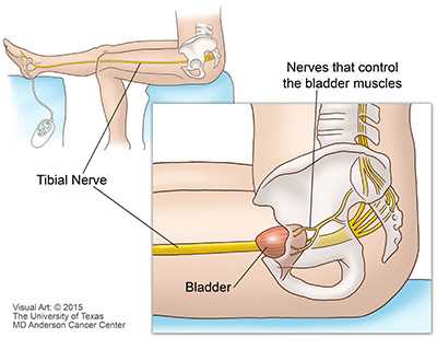
For effective diagnosis of urinary conditions, healthcare professionals often rely on direct observation techniques to assess the internal structures of the urinary system. This method allows specialists to identify abnormalities and detect issues that may not be visible through traditional imaging methods. By inspecting the interior of the system, a more accurate understanding of a patient’s health can be achieved.
Understanding these methods is crucial for anyone undergoing treatment or interested in urological care. These procedures can reveal various conditions, from infections to tumors, that might otherwise go unnoticed. Equipped with the right tools, doctors can make informed decisions about treatment options and monitor any changes over time.
Such assessments are essential for early detection and prevention of more serious health problems. Whether used for routine checkups or in response to specific symptoms, these diagnostic techniques play a vital role in maintaining long-term urinary health.
Visual Examination of the Bladder
Direct observation of internal urinary structures is an essential procedure in modern urology. It allows healthcare providers to inspect the organs responsible for urine storage and elimination. By accessing these areas, specialists can identify abnormal growths, blockages, or signs of infection that may affect a patient’s health. This approach provides a clear view of any potential issues, enabling early intervention and more precise treatment planning.
Techniques Used for Internal Observation
One of the most commonly used methods for internal visualization is cystoscopy, which involves inserting a thin tube with a camera into the urinary tract. This tool allows specialists to examine the inner lining of the organs in real-time. Other methods may include imaging techniques or less invasive scopes that provide crucial insight without the need for surgery. Each technique has its benefits depending on the patient’s symptoms and medical history.
Conditions Identified Through Direct Inspection
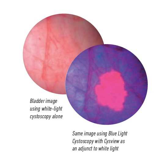
Through internal observation, healthcare professionals can detect a wide range of conditions, including infections, tumors, or signs of inflammation. Early detection is vital for successful treatment, as many issues can worsen without intervention. Additionally, this approach can be used for ongoing monitoring of chronic conditions, ensuring that any changes in the urinary system are promptly addressed.
Understanding Bladder Health Through Visual Exam
Maintaining urinary system health is crucial for overall well-being. Direct observation methods provide detailed insights into the functioning of organs responsible for storing and eliminating urine. These procedures help identify a variety of conditions that may not be noticeable through other diagnostic techniques, offering a clearer view of internal health. By monitoring the state of these organs, healthcare providers can ensure early detection and more effective treatment of potential issues.
Several key conditions can be identified through this method, including:
- Infections that cause discomfort or urinary problems.
- Tumors or abnormal growths that might indicate cancer.
- Blockages that interfere with normal urine flow.
- Inflammation or damage to the inner tissues of the system.
Early detection of such conditions is critical, as it allows for prompt intervention, preventing complications that could lead to more severe health issues. These procedures are particularly valuable in diagnosing chronic conditions that may require ongoing management and monitoring.
Role of Visual Inspection in Urinary Diagnosis

Direct observation methods play a pivotal role in diagnosing urinary system disorders. By providing a clear view of internal structures, these techniques allow healthcare professionals to assess the health of organs responsible for urine storage and elimination. They offer a detailed perspective on potential abnormalities that may not be detected through conventional imaging or lab tests, making them essential for accurate diagnosis and treatment planning.
Below is a table illustrating how inspection contributes to the identification of various conditions in the urinary system:
| Condition | How It’s Identified | Potential Impact |
|---|---|---|
| Infections | Inflammation or abnormal findings in tissues | Can cause pain, frequent urination, and discomfort |
| Obstructions | Detection of blockages or narrowing of passages | Disrupts urine flow, may lead to kidney damage |
| Growths or Tumors | Visible abnormal growths in organ lining | May indicate cancer or benign conditions requiring treatment |
| Hematuria | Presence of blood in urine or visual signs of bleeding | Could be a sign of infection, stones, or cancer |
With early detection, many of these conditions can be managed effectively, improving patient outcomes and preventing more serious health complications. The role of direct inspection cannot be overstated, as it aids in diagnosing and monitoring ongoing issues that affect urinary system health.
Methods Used for Bladder Visualization
Various techniques are employed to closely inspect the internal structures of the urinary system. These methods allow healthcare providers to observe potential abnormalities, diagnose conditions, and monitor ongoing treatment. The choice of method often depends on the patient’s symptoms, medical history, and the specific area requiring assessment.
Cystoscopy
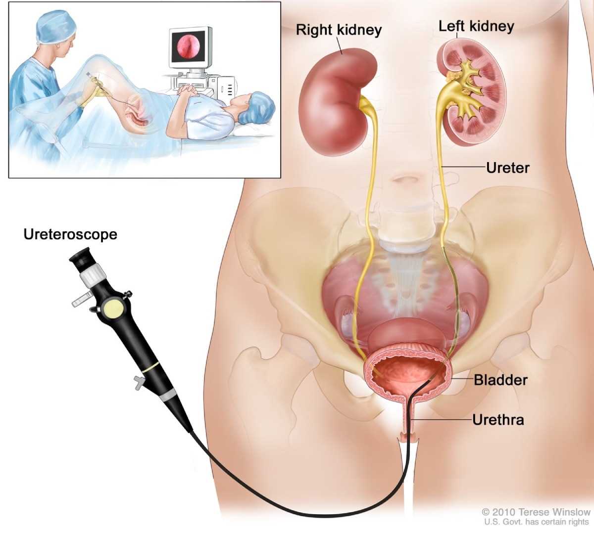
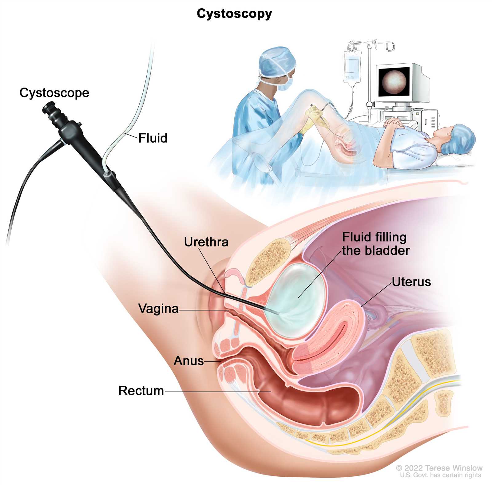
Cystoscopy is one of the most common procedures for internal inspection. A thin tube with a light and camera is inserted into the urinary tract, offering a real-time view of the organs. This approach is highly effective for diagnosing conditions such as infections, tumors, or blockages. Cystoscopy can also be used for therapeutic purposes, such as removing small stones or taking tissue samples for biopsy.
Ultrasound Imaging
Ultrasound imaging provides a non-invasive method of examining the urinary system. High-frequency sound waves create detailed images of the organs without the need for any incisions or tubes. It is particularly useful for detecting abnormalities such as kidney stones, tumors, and bladder wall changes. While not as detailed as cystoscopy, ultrasound is often used as an initial diagnostic tool.
These methods are complemented by other imaging techniques, such as MRI and CT scans, which provide additional insights for complex cases or when a more thorough examination is needed. Each method has its own strengths, offering healthcare providers a comprehensive toolkit for accurate diagnosis and effective treatment.
How a Cystoscopy Examines the Bladder
Cystoscopy is a key procedure used to inspect the interior of the urinary system. It involves using a specialized tool that allows healthcare professionals to directly view the organs involved in urine storage and elimination. This method provides real-time images, helping specialists identify abnormalities such as growths, blockages, or signs of inflammation.
Steps Involved in the Procedure
During a cystoscopy, the following steps are typically involved:
- Preparation: The patient is positioned comfortably, and local anesthesia is applied to minimize discomfort.
- Insertion of the Scope: A thin, flexible tube with a camera is gently inserted into the urinary tract through the urethra.
- Inspection: The scope sends images to a monitor, allowing the healthcare provider to examine the organs closely.
- Completion: After the procedure, the scope is carefully removed, and the patient is monitored for any immediate discomfort or complications.
Conditions Diagnosed with Cystoscopy

Several conditions can be detected through cystoscopy, including:
- Infections: Signs of inflammation or infection within the system.
- Growths: Tumors, polyps, or abnormal tissue growths that may require biopsy or removal.
- Blockages: Obstructions that hinder normal urine flow, which may cause discomfort or more severe complications.
- Damage: Injuries or scars that may have occurred due to past procedures or chronic conditions.
This procedure is minimally invasive and provides highly accurate results, making it an essential tool for diagnosing various urinary tract disorders. It also offers the possibility for treatment during the same procedure, such as removing small stones or taking tissue samples for further analysis.
Importance of Visual Exam in Urology
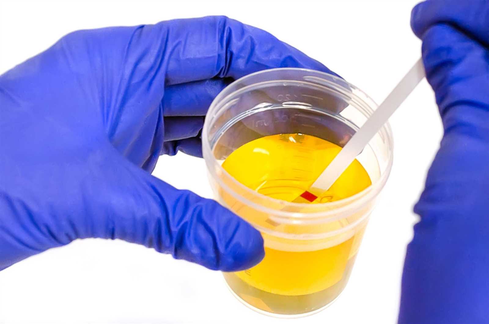
Direct observation techniques are essential in urology for diagnosing and managing various conditions affecting the urinary system. These methods provide real-time insights into the internal structures, allowing specialists to detect abnormalities, infections, and other issues that might not be visible through traditional tests. By offering a closer look at these organs, these procedures help in formulating accurate diagnoses and effective treatment plans.
Some of the key reasons for incorporating visual assessments into urological practice include:
- Early Detection: Identifying issues such as tumors, infections, or obstructions before they become severe.
- Precision in Diagnosis: Confirming diagnoses with high accuracy based on real-time observations.
- Monitoring Chronic Conditions: Keeping track of ongoing health conditions and ensuring they are managed effectively.
- Minimally Invasive Treatment: Performing certain therapeutic procedures during the same session, such as removing small stones or taking biopsies.
These techniques also play a crucial role in patient care, offering a less invasive and more efficient way to evaluate the urinary system compared to traditional surgical methods. With their ability to detect problems early, they significantly contribute to improving patient outcomes and reducing the need for more complex interventions.
Detecting Abnormalities During Bladder Inspection
During internal examinations of the urinary system, healthcare professionals can identify a variety of abnormal conditions that may not be detected through other diagnostic methods. These procedures provide a direct view of organs involved in urine storage and elimination, allowing specialists to spot issues such as growths, inflammation, or blockages. Early identification of these abnormalities is crucial for effective treatment and prevention of more serious health problems.
Common conditions that can be detected include:
- Tumors and Growths: Abnormal tissue growths, including benign polyps or malignant tumors, that may require further analysis or removal.
- Infections: Signs of inflammation or infection, such as redness, swelling, or pus, that can indicate bacterial or viral infections within the urinary system.
- Obstructions: Blockages in the urethra or other parts of the system that can affect the flow of urine, leading to discomfort or damage.
- Scarring and Damage: Evidence of past injuries or chronic conditions that have caused changes to the inner walls of organs, leading to potential functional issues.
- Hematuria: Blood in the urine or visible signs of bleeding, which could indicate a variety of underlying issues such as stones, infection, or cancer.
By detecting these abnormalities early, specialists can take appropriate steps to address the issue, whether through medication, minimally invasive procedures, or further testing. Regular inspections can help prevent conditions from escalating into more severe health problems, improving overall patient outcomes.
Bladder Cancer Detection Through Visual Methods
Detecting cancerous growths in organs responsible for urine storage is crucial for early intervention and treatment. Direct observation methods play a vital role in identifying abnormal tissue or tumors that could be indicative of malignancy. These procedures allow healthcare professionals to examine the inner surfaces of organs and recognize suspicious changes that might otherwise go unnoticed through other diagnostic tests.
One of the most effective techniques for identifying cancer in the urinary system is through a direct internal inspection. Here’s a comparison of various methods used to detect cancerous cells:
| Method | Detection Process | Effectiveness |
|---|---|---|
| Cystoscopy | Camera-equipped tube inserted to observe the inner lining of urinary organs, detecting tumors or abnormal growths. | Highly effective, providing clear images for diagnosis and biopsy. |
| Urinary Cytology | Microscopic examination of urine samples to detect cancerous cells. | Useful as a complementary tool, though less specific than direct visualization. |
| Ultrasound | High-frequency sound waves create images of internal structures, spotting larger tumors or irregularities. | Effective for initial detection but may not capture smaller growths. |
Early detection of cancer through these methods enables timely treatment, improving patient survival rates. Whether through biopsy, removal of tissue samples, or further imaging, these techniques remain essential in diagnosing and managing urinary tract cancers.
Preparation for a Bladder Visual Exam
Proper preparation is essential for ensuring that internal inspections of the urinary system are accurate and effective. Preparing for these procedures involves a combination of patient education, physical readiness, and medical evaluation. It ensures the process is as smooth as possible, minimizing discomfort and improving the likelihood of obtaining clear, reliable results.
Patient Instructions
Patients are typically given specific instructions to follow in advance of the procedure. These may include:
- Fasting: Some procedures may require the patient to avoid eating or drinking for several hours beforehand to ensure the organs are visible and unobstructed.
- Medication Adjustments: Certain medications, especially blood thinners or diuretics, may need to be paused or adjusted based on the doctor’s recommendations.
- Hydration: In some cases, patients are instructed to drink plenty of water before the procedure to ensure a full bladder, which can improve visibility during the inspection.
Physical and Emotional Preparation
In addition to medical instructions, patients may also be guided to prepare emotionally for the procedure. Understanding the steps involved can reduce anxiety and help individuals feel more comfortable with the process. Healthcare providers should explain:
- Procedure Duration: Most procedures are quick and non-invasive, but knowing what to expect can alleviate any nervousness.
- Possible Discomfort: While generally well-tolerated, some mild discomfort or pressure may occur during the insertion of instruments, which can be alleviated by anesthesia or pain management techniques.
By following these instructions and preparing mentally, patients are more likely to experience a successful and less stressful procedure, leading to more accurate diagnostic results.
What to Expect During a Bladder Exam
During an internal assessment of the urinary system, patients may feel some uncertainty about what will occur. This procedure is typically straightforward, with healthcare professionals guiding the patient through each step to ensure comfort and clarity. The main goal is to observe the inner surfaces of urinary organs for any signs of abnormality or disease.
Initially, the patient will be positioned in a comfortable manner, typically lying down, with easy access to the relevant areas. A local anesthetic or mild sedative may be administered to minimize discomfort, depending on the procedure. The healthcare provider will then introduce a specialized instrument to gently navigate through the urinary tract for an up-close look at the internal structures.
During the process, patients may experience slight pressure or a sensation of fullness, especially if the procedure involves viewing a distended organ. It’s important to communicate any discomfort with the medical team, who will adjust as needed to ensure minimal distress. Most procedures are brief, lasting only a few minutes, and do not require overnight stays in a medical facility.
After the procedure, patients can generally resume normal activities, although they may be advised to avoid strenuous activities for a short period. Depending on the findings, further testing or treatments may be recommended to address any concerns uncovered during the assessment.
Common Conditions Identified in Bladder Visual Exams

During internal assessments of the urinary system, a variety of conditions can be identified, ranging from minor irritations to more serious health concerns. These procedures provide a direct view of the inner structures, allowing medical professionals to detect issues that may not be visible through other diagnostic methods. Some of the most common conditions that may be discovered include:
- Infections: Inflammation or infection within the urinary tract is common, often presenting as redness, swelling, or visible discharge. These can be caused by bacterial or viral agents.
- Polyps and Tumors: Abnormal growths, including benign polyps or malignant tumors, can be detected. These may require further testing, such as biopsies, to determine their nature and treatment options.
- Urinary Stones: Hard deposits of minerals can form within the urinary organs, causing blockages, pain, or difficulty urinating. Stones are often identified during internal observations and can vary in size.
- Hematuria: The presence of blood in urine may be seen, which can indicate a variety of underlying issues such as trauma, infections, or even cancerous growths.
- Scarring: Scarring from previous infections, surgeries, or other conditions can alter the internal structure and function of the urinary organs, often leading to recurring issues.
- Diverticula: These are small pouches that can form in the walls of urinary organs. While often asymptomatic, they can sometimes lead to infections or complications.
- Bladder Diverticulitis: This is an infection of the diverticula, which can cause pain and discomfort. It can be identified through direct observation and treated with antibiotics or surgery.
Identifying these conditions early through internal assessment allows for timely treatment, minimizing the risk of complications and improving patient outcomes. Depending on the severity and type of condition, further diagnostic procedures or treatments may be required to address the issue appropriately.
Bladder Infections Seen Through Visual Inspection

Infections within the urinary tract can often be detected through internal assessments of the affected organs. These infections typically result in visible signs of inflammation, which can help healthcare providers identify the issue early. By observing the internal surfaces, healthcare professionals can spot indicators of infection, including redness, swelling, or unusual discharge. Early detection allows for timely treatment, reducing the risk of complications and promoting faster recovery.
During these assessments, the following signs of infection may be observed:
- Redness and Inflammation: Swollen tissue with a reddish hue often indicates irritation or infection caused by bacteria or viruses.
- Visible Pus or Discharge: Presence of yellow or greenish fluid suggests bacterial infection that may require antibiotics to clear up.
- Ulcers or Sores: Sores or open areas on the internal lining may form as a result of prolonged infection, making the area more vulnerable to further irritation.
- Foul Odor: An unpleasant odor during an internal observation can be indicative of an active infection or bacterial growth.
While these signs can point to an infection, further testing, such as urine culture or blood tests, may be necessary to identify the exact cause and determine the appropriate treatment. Proper diagnosis is crucial to addressing the infection effectively and preventing recurrence or spread to other areas.
Advantages of Visual Bladder Examination Techniques
Internal inspection methods offer numerous benefits in diagnosing and monitoring urinary system conditions. These techniques allow healthcare professionals to directly view the inner structures, providing valuable insights into any abnormalities or diseases that may be present. The ability to visualize the organs in real-time enhances diagnostic accuracy and aids in forming effective treatment plans.
Some of the key advantages include:
- Early Detection: The ability to directly observe internal conditions allows for the identification of issues at an early stage, often before symptoms are noticeable. This can lead to quicker treatment and better outcomes.
- Minimally Invasive: These methods are generally minimally invasive, requiring only small incisions or natural body openings for access, reducing recovery time and complications.
- Accurate Diagnosis: Direct visualization offers greater diagnostic precision, helping to differentiate between various conditions that might otherwise appear similar through imaging or lab tests alone.
- Real-time Monitoring: Physicians can monitor and assess any changes in the condition of the organs during the procedure, which is helpful in tracking disease progression or treatment effectiveness.
- Guided Biopsies and Treatments: When necessary, these techniques allow for targeted biopsies or treatments, reducing the risk of damage to surrounding tissue and ensuring a more focused approach to care.
- Reduced Need for Other Tests: Internal visualization may reduce the need for additional imaging or diagnostic tests, offering a more streamlined approach to patient care.
By providing clear and immediate access to internal structures, these techniques enhance both diagnostic confidence and treatment planning, ultimately improving patient care and outcomes.
Risks and Complications of Visual Bladder Exams
While internal assessments of the urinary system are crucial for accurate diagnosis, they do carry certain risks and potential complications. Though generally considered safe, complications can arise due to the invasive nature of the procedure, patient-specific factors, or technical difficulties during the procedure. Understanding these risks helps patients and healthcare providers make informed decisions and take necessary precautions.
Potential Risks
Some of the most common risks associated with these procedures include:
- Infection: Any internal procedure, even minimally invasive, can introduce bacteria, leading to urinary tract infections or more serious complications if not managed properly.
- Bleeding: In some cases, irritation of the tissues during the procedure may cause minor bleeding. While this is often temporary, excessive bleeding can require further intervention.
- Perforation: Though rare, accidental injury to the surrounding tissues or organs may occur, potentially leading to more severe complications and requiring surgical repair.
- Discomfort: Patients may experience temporary pain, burning, or discomfort during and after the procedure as the internal tissues recover.
Managing and Minimizing Risks
To minimize the likelihood of complications, healthcare providers follow strict protocols and guidelines. These include:
| Risk | Prevention/Management |
|---|---|
| Infection | Proper sterilization of equipment and prophylactic antibiotics may be administered to reduce the risk. |
| Bleeding | Careful technique and monitoring throughout the procedure ensure any bleeding is controlled quickly. |
| Perforation | Using advanced imaging technology during the procedure helps guide instruments to avoid injury to surrounding tissues. |
| Discomfort | Local anesthesia or mild sedation is often used to minimize discomfort and ease patient anxiety. |
While complications are possible, they are rare, and the benefits of early detection and accurate diagnosis generally outweigh the risks. By discussing potential concerns with healthcare providers, patients can better understand what to expect and how to prepare for a safe and effective procedure.
Post-Exam Care Following Bladder Inspection

After undergoing an internal evaluation of the urinary system, it is important to follow specific care instructions to promote healing and prevent complications. While most individuals recover quickly and experience minimal discomfort, certain precautions and post-procedure guidelines can help ensure a smooth recovery and prevent issues such as infection or irritation. Proper aftercare plays a key role in enhancing patient comfort and well-being during the recovery period.
General Care Instructions
After the procedure, patients may experience mild discomfort or irritation, but these effects are typically short-lived. Below are some general post-procedure recommendations:
- Hydration: It is essential to drink plenty of fluids following the procedure to help flush out any remaining contrast material and prevent urinary tract infections.
- Avoid Strenuous Activity: Patients should refrain from heavy physical activity or lifting for at least 24 hours to allow the urinary system time to heal.
- Rest: Adequate rest is important for recovery. Resting helps the body focus on healing and reduces the risk of complications.
- Manage Discomfort: Mild pain or discomfort can usually be controlled with over-the-counter pain relievers as recommended by the healthcare provider.
Potential Complications and Warning Signs

While rare, there are some warning signs to be aware of after the procedure. If any of the following symptoms occur, patients should contact their healthcare provider immediately:
| Symptom | Possible Concern | Action |
|---|---|---|
| Severe Pain | Possible injury or complications | Seek immediate medical attention |
| Fever | Infection | Contact healthcare provider |
| Heavy Bleeding | Excessive trauma to tissues | Go to the emergency room |
| Difficulty Urinating | Obstruction or irritation | Notify healthcare provider |
Following these guidelines and being aware of potential complications can significantly reduce the risk of issues after the procedure. By taking proper care and monitoring any symptoms, individuals can ensure a faster and safer recovery.
Comparing Visual Exams to Other Diagnostic Methods
When diagnosing conditions related to the urinary system, various techniques can be employed to gather information. Each method offers unique benefits and limitations depending on the patient’s symptoms, medical history, and the suspected condition. The internal inspection of the urinary tract provides a direct view of any abnormalities but may be complemented or even replaced by other diagnostic procedures, depending on the circumstances. Understanding the differences between these methods can help healthcare providers determine the best course of action for their patients.
Direct Observation vs. Imaging Techniques

Direct observation methods allow healthcare providers to visually examine internal structures and identify irregularities. However, imaging techniques, such as ultrasound, X-rays, or CT scans, are also frequently used. Below is a comparison of these methods:
- Direct Observation: Involves using a scope to view internal tissues directly, providing immediate results and the ability to take biopsies or samples during the procedure.
- Ultrasound: Non-invasive and commonly used for initial screenings. Provides images of soft tissues but lacks the detail needed for in-depth examination of internal structures.
- X-ray and CT Scans: Useful for identifying structural abnormalities or tumors, but less effective at detecting subtle changes or providing detailed tissue samples compared to direct visualization.
Non-Invasive Methods vs. Invasive Techniques
Non-invasive methods offer a less risky approach, but they may not always provide the level of detail needed for a definitive diagnosis. In contrast, invasive techniques such as direct observation are generally more effective for detecting specific issues.
- Non-invasive: Techniques like ultrasound, MRI, or urine tests are safe and painless but may not always pinpoint problems within internal structures with the same accuracy.
- Invasive: Direct internal inspection offers more accurate information for conditions such as tumors, stones, or inflammation. However, it involves some risk and recovery time for the patient.
In conclusion, while each diagnostic method has its strengths, choosing the most appropriate approach depends on the patient’s condition, the detail needed, and the available technology. A comprehensive diagnosis often involves a combination of techniques to ensure accuracy and provide the best care possible.
Future Innovations in Bladder Visual Examinations
As medical technology continues to evolve, new techniques and tools are emerging to enhance the accuracy and efficiency of diagnosing issues within the urinary tract. Innovations in imaging technologies, sensor development, and artificial intelligence are paving the way for more precise, non-invasive, and real-time assessments. These advancements aim to improve the overall patient experience while ensuring more detailed and earlier detection of abnormalities.
In the future, we can expect to see the integration of cutting-edge technologies such as advanced imaging devices, AI-assisted analysis, and improved endoscopic methods. These tools are designed to enhance diagnostic precision, reduce patient discomfort, and shorten recovery times. Below are some key innovations that are shaping the future of urinary system diagnostics:
- AI-Driven Analysis: Artificial intelligence can help process vast amounts of data from imaging devices, identifying patterns and abnormalities that may be difficult for human eyes to detect. This could lead to faster and more accurate diagnoses, improving outcomes for patients.
- High-Resolution Imaging: The development of high-resolution optical and infrared imaging technologies will provide unprecedented clarity, allowing for the detection of even the smallest anomalies within internal tissues.
- Non-Invasive Imaging Devices: New non-invasive technologies, such as advanced ultrasound or MRI techniques, could reduce the need for invasive procedures while still providing the detailed information required for diagnosis.
- Miniaturized Endoscopes: Smaller and more flexible endoscopic tools will enable better access to hard-to-reach areas, allowing for more detailed internal inspections without causing significant discomfort.
These innovations are expected to revolutionize how urinary system conditions are diagnosed and treated, leading to better patient care and improved clinical outcomes. As these technologies become more refined and accessible, they will likely play a critical role in shaping the future of urology and other medical fields.